

Anti-CEACAM1 [B3-17]
Ab01369-10.3
ApplicationsFlow Cytometry, ImmunoFluorescence, ImmunoPrecipitation, ELISA, ImmunoHistoChemistry
Product group Antibodies
TargetCEACAM1
Overview
- SupplierAbsolute Antibody
- Product NameAnti-CEACAM1 [B3-17]
- Delivery Days Customer7
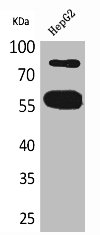
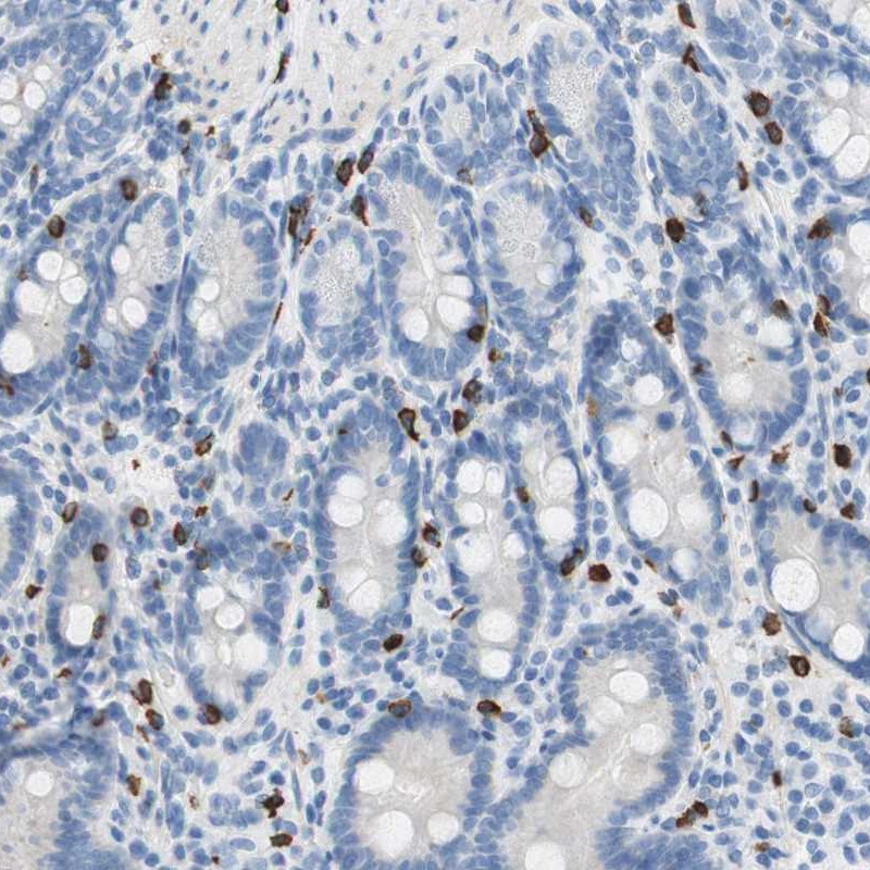
- Application Supplier NoteIn flow cytometric analysis, this antibody has been used to detect CEACAM1 surface expression by human epithelial and endothelial cell lines, and microvesicles derived from these cells (Muturi et al, 2013), normal human bronchial epithelial (NHBE) cells (Klaile et al, 2013), CEACAM1-knockout human intestinal epithelial cells (Klaile et al, 2017) and human B-cell subpopulations (Khairnar et al, 2015; Seifert et al, 2015). It has also been used to detect CEACAM1 expression in ELISA analysis of transfected A549 epithelial cells (Singer et al, 2014). This antibody has been used in immunofluorescence analysis of NHBE cells (Klaile et al, 2013), as well as in the immunoprecipitation of CEACAM1 from NHBE cell culture supernatants (Klaile et al, 2013). Additionally, this antibody has been used in IHC staining of paraffin-embedded human lung tissue sections (Klaile et al, 2013).
- ApplicationsFlow Cytometry, ImmunoFluorescence, ImmunoPrecipitation, ELISA, ImmunoHistoChemistry
- Applications SupplierIP; IHC; IF; FC; ELISA
- CertificationResearch Use Only
- ClonalityMonoclonal
- Clone IDB3-17
- Gene ID634
- Target nameCEACAM1
- Target descriptionCEA cell adhesion molecule 1
- Target synonymsBGP, BGP1, BGPI, cell adhesion molecule CEACAM1, CD66a antigen, antigen CD66, carcinoembryonic antigen related cell adhesion molecule 1, carcinoembryonic antigen-related cell adhesion molecule 1 (biliary glycoprotein)
- HostHuman
- IsotypeIgG1
- Protein IDP13688
- Protein NameCarcinoembryonic antigen-related cell adhesion molecule 1
- Scientific DescriptionThis chimeric human antibody was made using the variable domain sequences of the original Mouse IgG1 format, for improved compatibility with existing reagents, assays and techniques.
- Reactivity SupplierHuman
- Reactivity Supplier NoteThis antibody was raised by immunising mice with recombinant human CEACAM1.
- Storage Instruction-20°C,2°C to 8°C
- UNSPSC12352203
Related products
Product group Antibodies
CEACAM1 AntibodyCSB-PA005134
ApplicationsWestern Blot, ELISA
ReactivityHuman
TargetCEACAM1
- SizePrice
Product group Antibodies
CEACAM1 antibody [N1N3]GTX113392
ApplicationsWestern Blot, ImmunoHistoChemistry, ImmunoHistoChemistry Paraffin
TargetCEACAM1
- SizePrice
Product group Antibodies
ApplicationsFlow Cytometry, ImmunoPrecipitation, Western Blot, ImmunoCytoChemistry, ImmunoHistoChemistry
TargetCEACAM1
- SizePrice
Product group Antibodies

Anti-CD66acd [YTH 71.3]Ab00198-23.0
ApplicationsFlow Cytometry, ImmunoHistoChemistry, ImmunoHistoChemistry Frozen
TargetCEACAM1
- SizePrice
Product group Antibodies
Anti-CEACAM1 Antibody144-01702
ApplicationsWestern Blot
TargetCEACAM1
- SizePrice
Product group Antibodies
Anti-CEACAM1 AntibodyHPA011041
ApplicationsImmunoHistoChemistry
ReactivityHuman
TargetCEACAM1
- SizePrice

Product group Antibodies
Anti-CEACAM1 Antibody Picoband(r)A00923-2-CARRIER-FREE
ApplicationsWestern Blot, ELISA
TargetCEACAM1
- SizePrice
Product group Antibodies
CEACAM1 Polyclonal AntibodyCAC14123
ApplicationsImmunoFluorescence, Western Blot, ELISA
TargetCEACAM1
- SizePrice