

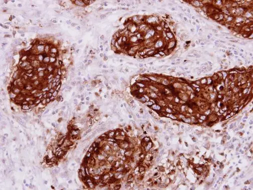
C-CAM1 antibody [N1N3] detects C-CAM1 protein at cytoplasm on human colon carcinoma by immunohistochemical analysis. Sample: Paraffin-embedded colon carcinoma. C-CAM1 antibody [N1N3] (GTX113392) dilution: 1:250.
Antigen Retrieval: Trilogy? (EDTA based, pH 8.0) buffer, 15min
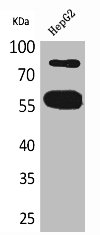
![Human colon (30 μg) was separated by 5% SDS-PAGE, and the membrane was blotted with CEACAM1 antibody [N1N3] (GTX113392) diluted at 1:500. The HRP-conjugated anti-rabbit IgG antibody (GTX213110-01) was used to detect the primary antibody.](https://www.genetex.com/upload/website/prouct_img/normal/GTX113392/GTX113392_40100_20240202_WB_colon_24021917_208.webp "Human colon (30 μg) was separated by 5% SDS-PAGE, and the membrane was blotted with CEACAM1 antibody [N1N3] (GTX113392) diluted at 1:500. The HRP-conjugated anti-rabbit IgG antibody (GTX213110-01) was used to detect the primary antibody.")
C-CAM1 antibody [N1N3] detects C-CAM1 protein at cytoplasm on human colon carcinoma by immunohistochemical analysis. Sample: Paraffin-embedded colon carcinoma. C-CAM1 antibody [N1N3] (GTX113392) dilution: 1:250.
Antigen Retrieval: Trilogy? (EDTA based, pH 8.0) buffer, 15min
CEACAM1 antibody [N1N3]
GTX113392
ApplicationsWestern Blot, ImmunoHistoChemistry, ImmunoHistoChemistry Paraffin
Product group Antibodies
ReactivityHuman
TargetCEACAM1
Overview
- SupplierGeneTex
- Product NameCEACAM1 antibody [N1N3]
- Delivery Days Customer9
- Application Supplier NoteWB: 1:500-1:3000. IHC-P: 1:100-1:1000. *Optimal dilutions/concentrations should be determined by the researcher.Not tested in other applications.
- ApplicationsWestern Blot, ImmunoHistoChemistry, ImmunoHistoChemistry Paraffin
- CertificationResearch Use Only
- ClonalityPolyclonal
- Concentration1 mg/ml
- ConjugateUnconjugated
- Gene ID634
- Target nameCEACAM1
- Target descriptionCEA cell adhesion molecule 1
- Target synonymsBGP, BGP1, BGPI, cell adhesion molecule CEACAM1, CD66a antigen, antigen CD66, carcinoembryonic antigen related cell adhesion molecule 1, carcinoembryonic antigen-related cell adhesion molecule 1 (biliary glycoprotein)
- HostRabbit
- IsotypeIgG
- Protein IDP13688
- Protein NameCell adhesion molecule CEACAM1
- Scientific DescriptionThis gene encodes a member of the carcinoembryonic antigen (CEA) gene family, which belongs to the immunoglobulin superfamily. Two subgroups of the CEA family, the CEA cell adhesion molecules and the pregnancy-specific glycoproteins, are located within a 1.2 Mb cluster on the long arm of chromosome 19. Eleven pseudogenes of the CEA cell adhesion molecule subgroup are also found in the cluster. The encoded protein was originally described in bile ducts of liver as biliary glycoprotein. Subsequently, it was found to be a cell-cell adhesion molecule detected on leukocytes, epithelia, and endothelia. The encoded protein mediates cell adhesion via homophilic as well as heterophilic binding to other proteins of the subgroup. Multiple cellular activities have been attributed to the encoded protein, including roles in the differentiation and arrangement of tissue three-dimensional structure, angiogenesis, apoptosis, tumor suppression, metastasis, and the modulation of innate and adaptive immune responses. Multiple transcript variants encoding different isoforms have been reported, but the full-length nature of only two has been determined. [provided by RefSeq]
- ReactivityHuman
- Storage Instruction-20°C or -80°C,2°C to 8°C
- UNSPSC41116161
Datasheet
Related products
Product group Antibodies
Anti-CEACAM1 AntibodyA31625
ApplicationsWestern Blot, ImmunoHistoChemistry
ReactivityHuman, Mouse
- SizePrice
Product group Antibodies

Anti-CD66acd [YTH 71.3]Ab00198-23.0
ApplicationsFlow Cytometry, ImmunoHistoChemistry, ImmunoHistoChemistry Frozen
ReactivityHuman, Primate
TargetCEACAM1
- SizePrice

Product group Antibodies
Anti-CEACAM1 Antibody Picoband(r)A00923-2-CARRIER-FREE
ApplicationsWestern Blot, ELISA
ReactivityHuman
TargetCEACAM1
- SizePrice
Product group Antibodies
Anti-CEACAM1 Antibody144-01702
ApplicationsWestern Blot
ReactivityHuman, Mouse, Rat
TargetCEACAM1
- SizePrice
Product group Antibodies

CD66a / CEACAM1 AntibodyLS-C831499
ApplicationsImmunoHistoChemistry
ReactivityMouse, Rat
TargetCEACAM1
- SizePrice
Product group Antibodies
CD66a Recombinant AntibodyBSM-60348R
ApplicationsImmunoFluorescence, Western Blot, ImmunoHistoChemistry, ImmunoHistoChemistry Frozen, ImmunoHistoChemistry Paraffin
ReactivityHuman
TargetCEACAM1
- SizePrice
Product group Antibodies
CEACAM1 Polyclonal AntibodyCAC14123
ApplicationsImmunoFluorescence, Western Blot, ELISA
TargetCEACAM1
- SizePrice
Product group Antibodies
CEACAM1 AntibodyCSB-PA005134
ApplicationsWestern Blot, ELISA
ReactivityHuman
TargetCEACAM1
- SizePrice
Product group Antibodies
Anti-CEACAM1 AntibodyHPA011041
ApplicationsImmunoHistoChemistry
ReactivityHuman
TargetCEACAM1
- SizePrice
Product group Antibodies
CEACAM1 antibody [B464M]GTX44154
ApplicationsELISA, ImmunoHistoChemistry
ReactivityHuman
TargetCEACAM1
- SizePrice