


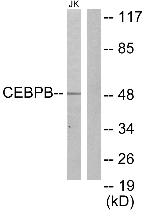
Figure 1. Western blot analysis of CEBP Beta using anti-CEBP Beta antibody (PB9171). Electrophoresis was performed on a 5-20% SDS-PAGE gel at 70V (Stacking gel) / 90V (Resolving gel) for 2-3 hours. The sample well of each lane was loaded with 30 ug of sample under reducing conditions. Lane 1: human Hela whole cell lysates, Lane 2: human U87 whole cell lysates. After electrophoresis, proteins were transferred to a nitrocellulose membrane at 150 mA for 50-90 minutes. Blocked the membrane with 5% non-fat milk/TBS for 1.5 hour at RT. The membrane was incubated with rabbit anti-CEBP Beta antigen affinity purified polyclonal antibody (Catalog # PB9171) at 0.5 microg/mL overnight at 4°C, then washed with TBS-0.1%Tween 3 times with 5 minutes each and probed with a goat anti-rabbit IgG-HRP secondary antibody at a dilution of 1:5000 for 1.5 hour at RT. The signal is developed using an Enhanced Chemiluminescent detection (ECL) kit (Catalog # EK1002) with Tanon 5200 system. A specific band was detected for CEBP Beta at approximately 42, 46 kDa. The expected band size for CEBP Beta is at 36 kDa.
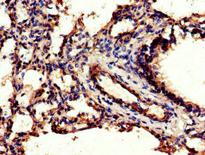
. CEBP Beta was detected in a paraffin-embedded section of human placenta tissue. Heat mediated antigen retrieval was performed in EDTA buffer (pH 8.0, epitope retrieval solution). The tissue section was blocked with 10% goat serum. The tissue section was then incubated with 2 microg/ml rabbit anti-CEBP Beta Antibody (PB9171) overnight at 4°C. Peroxidase Conjugated Goat Anti-rabbit IgG was used as secondary antibody and incubated for 30 minutes at 37°C. The tissue section was developed using HRP Conjugated Rabbit IgG Super Vision Assay Kit (Catalog # SV0002) with DAB as the chromogen.")
. CEBP Beta was detected in a paraffin-embedded section of human squamous metaplasia of renal pelvis tissue. Heat mediated antigen retrieval was performed in EDTA buffer (pH 8.0, epitope retrieval solution). The tissue section was blocked with 10% goat serum. The tissue section was then incubated with 2 microg/ml rabbit anti-CEBP Beta Antibody (PB9171) overnight at 4°C. Peroxidase Conjugated Goat Anti-rabbit IgG was used as secondary antibody and incubated for 30 minutes at 37°C. The tissue section was developed using HRP Conjugated Rabbit IgG Super Vision Assay Kit (Catalog # SV0002) with DAB as the chromogen.")
. CEBP Beta was detected in immunocytochemical section of A431 cells. Enzyme antigen retrieval was performed using IHC enzyme antigen retrieval reagent (AR0022) for 15 mins. The cells were blocked with 10% goat serum. And then incubated with 5microg/mL rabbit anti-CEBP Beta Antibody (PB9171) overnight at 4°C. DyLight®488 Conjugated Goat Anti-Rabbit IgG (BA1127) was used as secondary antibody at 1:500 dilution and incubated for 30 minutes at 37°C. The section was counterstained with DAPI. Visualize using a fluorescence microscope and filter sets appropriate for the label used.")
. Overlay histogram showing A431 cells stained with PB9171 (Blue line). To facilitate intracellular staining, cells were fixed with 4% paraformaldehyde and permeabilized with permeabilization buffer. The cells were blocked with 10% normal goat serum. And then incubated with rabbit anti-CEBP Beta Antibody (PB9171, 1 microg/1x106 cells) for 30 min at 20°C. DyLight®488 conjugated goat anti-rabbit IgG (BA1127, 5-10 microg/1x106 cells) was used as secondary antibody for 30 minutes at 20°C. Isotype control antibody (Green line) was rabbit IgG (1 microg/1x106) used under the same conditions. Unlabelled sample without incubation with primary antibody and secondary antibody (Red line) was used as a blank control.")
Figure 1. Western blot analysis of CEBP Beta using anti-CEBP Beta antibody (PB9171). Electrophoresis was performed on a 5-20% SDS-PAGE gel at 70V (Stacking gel) / 90V (Resolving gel) for 2-3 hours. The sample well of each lane was loaded with 30 ug of sample under reducing conditions. Lane 1: human Hela whole cell lysates, Lane 2: human U87 whole cell lysates. After electrophoresis, proteins were transferred to a nitrocellulose membrane at 150 mA for 50-90 minutes. Blocked the membrane with 5% non-fat milk/TBS for 1.5 hour at RT. The membrane was incubated with rabbit anti-CEBP Beta antigen affinity purified polyclonal antibody (Catalog # PB9171) at 0.5 microg/mL overnight at 4°C, then washed with TBS-0.1%Tween 3 times with 5 minutes each and probed with a goat anti-rabbit IgG-HRP secondary antibody at a dilution of 1:5000 for 1.5 hour at RT. The signal is developed using an Enhanced Chemiluminescent detection (ECL) kit (Catalog # EK1002) with Tanon 5200 system. A specific band was detected for CEBP Beta at approximately 42, 46 kDa. The expected band size for CEBP Beta is at 36 kDa.
Anti-CEBP Beta/CEBPB Antibody Picoband(r)
PB9171
ApplicationsFlow Cytometry, ImmunoFluorescence, Western Blot, ImmunoCytoChemistry, ImmunoHistoChemistry
Product group Antibodies
ReactivityHuman
TargetCEBPB
Overview
- SupplierBoster Bio
- Product NameAnti-CEBP Beta/CEBPB Antibody Picoband(r)
- Delivery Days Customer9
- Application Supplier NoteWB: The detection limit for CEBP Beta is approximately 0.25ng/lane under reducing conditions. Tested Species: In-house tested species with positive results. By Heat: Boiling the paraffin sections in 10mM citrate buffer, pH6.0, for 20mins is required for the staining of formalin/paraffin sections. Other applications have not been tested. Optimal dilutions should be determined by end users.
- ApplicationsFlow Cytometry, ImmunoFluorescence, Western Blot, ImmunoCytoChemistry, ImmunoHistoChemistry
- Applications SupplierIHP, IHF, WB, IHC
- CertificationResearch Use Only
- ClonalityPolyclonal
- Concentration500 ug/ml
- Gene ID1051
- Target nameCEBPB
- Target descriptionCCAAT enhancer binding protein beta
- Target synonymsC/EBP-beta, IL6DBP, NF-IL6, TCF5, CCAAT/enhancer-binding protein beta, CCAAT/enhancer binding protein (C/EBP), beta, interleukin 6-dependent DNA-binding protein, nuclear factor NF-IL6, nuclear factor of interleukin 6, transcription factor 5, transcription factor C/EBP beta
- HostRabbit
- IsotypeIgG
- Protein IDP17676
- Protein NameCCAAT/enhancer-binding protein beta
- Scientific DescriptionBoster Bio Anti-CEBP Beta/CEBPB Antibody Picoband® catalog # PB9171. Tested in Flow Cytometry, IF, IHC, ICC, WB applications. This antibody reacts with Human. The brand Picoband indicates this is a premium antibody that guarantees superior quality, high affinity, and strong signals with minimal background in Western blot applications. Only our best-performing antibodies are designated as Picoband, ensuring unmatched performance.
- ReactivityHuman
- Reactivity SupplierHuman, Mouse, Rat
- Storage Instruction-20°C,2°C to 8°C
- UNSPSC12352203
References
- Liu Q, Zhao Y, Wang Q, et al. Convergent alteration of the mesenchymal stem cell heterogeneity in adipose tissue during aging. FASEB J. 2023,37(8):e23114. doi: 10.1096/fj.202300807RRead this paper
- Wang N, Li Y, Li Z, et al. Sal B targets TAZ to facilitate osteogenesis and reduce adipogenesis through MEK-ERK pathway. J Cell Mol Med. 2019,23(5):3683-3695. doi: 10.1111/jcmm.14272Read this paper
- Wang N, Li Y, Li Z, et al. IRS-1 targets TAZ to inhibit adipogenesis of rat bone marrow mesenchymal stem cells through PI3K-Akt and MEK-ERK pathways. Eur J Pharmacol. 2019,849:11-21. doi: 10.1016/j.ejphar.2019.01.064Read this paper
Datasheet
MSDS
Related products
Product group Antibodies
Anti-CEBPB AntibodyA99145
ApplicationsWestern Blot, ELISA
ReactivityHuman, Mouse
- SizePrice
Product group Antibodies
Anti-CEBPB Antibody130-10238
ApplicationsWestern Blot, ELISA
TargetCEBPB
- SizePrice
Product group Antibodies

ApplicationsWestern Blot, ELISA
ReactivityHuman
TargetCEBPB
- SizePrice
Product group Antibodies
CEBP beta Recombinant Antibody, Biotin ConjugatedBSM-61530R-BIOTIN
ApplicationsImmunoPrecipitation, Western Blot
ReactivityHuman, Mouse, Rat
TargetCEBPB
- SizePrice
Product group Antibodies

Goat anti-CEBPB (aa68-81)EB12113
ApplicationsWestern Blot, ELISA
ReactivityBovine, Canine, Human, Mouse, Porcine, Rat
TargetCEBPB
- SizePrice
Product group Antibodies
CEBPB AntibodyCSB-PA005181LA01HU
ApplicationsELISA, ImmunoHistoChemistry
ReactivityHuman
TargetCEBPB
- SizePrice
![C/EBP beta antibody detects C/EBP beta protein at cytoplasm and nucleus by immunofluorescent analysis. Sample: HeLa cells were fixed in 4% paraformaldehyde at RT for 15 min. Green: C/EBP beta stained by C/EBP beta antibody (GTX100675) diluted at 1:500. Red: alpha Tubulin, a cytoskeleton marker, stained by alpha Tubulin antibody [GT114] (GTX628802) diluted at 1:1000.](https://www.genetex.com/upload/website/prouct_img/normal/GTX100675/GTX100675_44664_20220624_ICC_IF_22062919_578.webp)
Product group Antibodies
C/EBP beta antibodyGTX100675
ApplicationsImmunoFluorescence, ImmunoPrecipitation, Western Blot, ChIP Chromatin ImmunoPrecipitation, ImmunoCytoChemistry, ImmunoHistoChemistry, ImmunoHistoChemistry Paraffin
ReactivityHuman
TargetCEBPB
- SizePrice

Product group Antibodies
Anti-CEBPB AntibodyHPA061355
ApplicationsWestern Blot, ImmunoCytoChemistry
ReactivityHuman
TargetCEBPB
- SizePrice
Product group Antibodies
Anti-CEBP Beta/CEBPB Antibody Picoband(r)PB9171-CARRIER-FREE
ApplicationsFlow Cytometry, ImmunoFluorescence, Western Blot, ImmunoCytoChemistry, ImmunoHistoChemistry
ReactivityHuman
TargetCEBPB
- SizePrice