


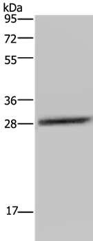
Figure 1. Western blot analysis of CEBP Delta/CEBPD using anti-CEBP Delta/CEBPD antibody (A03499-4). Electrophoresis was performed on a 5-20% SDS-PAGE gel at 70V (Stacking gel) / 90V (Resolving gel) for 2-3 hours. The sample well of each lane was loaded with 50ug of sample under reducing conditions. Lane 1: human A549 whole cell lysates, Lane 2: human placenta tissue lysates, Lane 3: human U87 whole cell lysates, Lane 4: mouse RAW264.7 whole cell lysates. After Electrophoresis, proteins were transferred to a Nitrocellulose membrane at 150mA for 50-90 minutes. Blocked the membrane with 5% Non-fat Milk/ TBS for 1.5 hour at RT. The membrane was incubated with rabbit anti-CEBP Delta/CEBPD antigen affinity purified polyclonal antibody (Catalog # A03499-4) at 0.5 microg/mL overnight at 4°C, then washed with TBS-0.1%Tween 3 times with 5 minutes each and probed with a goat anti-rabbit IgG-HRP secondary antibody at a dilution of 1:5000 for 1.5 hour at RT. The signal is developed using an Enhanced Chemiluminescent detection (ECL) kit (Catalog # EK1002) with Tanon 5200 system. A specific band was detected for CEBP Delta/CEBPD at approximately 36KD. The expected band size for CEBP Delta/CEBPD is at 36KD.
. Overlay histogram showing SiHa cells stained with A03499-4 (Blue line). To facilitate intracellular staining, cells were fixed with 4% paraformaldehyde and permeabilized with permeabilization buffer. The cells were blocked with 10% normal goat serum. And then incubated with rabbit anti-CEBP Delta/CEBPD Antibody (A03499-4, 1microg/1x106 cells) for 30 min at 20°C. DyLight®488 conjugated goat anti-rabbit IgG (BA1127, 5-10microg/1x106 cells) was used as secondary antibody for 30 minutes at 20°C. Isotype control antibody (Green line) was rabbit IgG (1microg/1x106) used under the same conditions. Unlabelled sample without incubation with primary antibody and secondary antibody (Red line) was used as a blank control.")
Figure 1. Western blot analysis of CEBP Delta/CEBPD using anti-CEBP Delta/CEBPD antibody (A03499-4). Electrophoresis was performed on a 5-20% SDS-PAGE gel at 70V (Stacking gel) / 90V (Resolving gel) for 2-3 hours. The sample well of each lane was loaded with 50ug of sample under reducing conditions. Lane 1: human A549 whole cell lysates, Lane 2: human placenta tissue lysates, Lane 3: human U87 whole cell lysates, Lane 4: mouse RAW264.7 whole cell lysates. After Electrophoresis, proteins were transferred to a Nitrocellulose membrane at 150mA for 50-90 minutes. Blocked the membrane with 5% Non-fat Milk/ TBS for 1.5 hour at RT. The membrane was incubated with rabbit anti-CEBP Delta/CEBPD antigen affinity purified polyclonal antibody (Catalog # A03499-4) at 0.5 microg/mL overnight at 4°C, then washed with TBS-0.1%Tween 3 times with 5 minutes each and probed with a goat anti-rabbit IgG-HRP secondary antibody at a dilution of 1:5000 for 1.5 hour at RT. The signal is developed using an Enhanced Chemiluminescent detection (ECL) kit (Catalog # EK1002) with Tanon 5200 system. A specific band was detected for CEBP Delta/CEBPD at approximately 36KD. The expected band size for CEBP Delta/CEBPD is at 36KD.
Anti-CEBP Delta/CEBPD Antibody Picoband(r)
A03499-4-CARRIER-FREE
ApplicationsFlow Cytometry, Western Blot, ELISA
Product group Antibodies
ReactivityHuman, Mouse
TargetCEBPD
Overview
- SupplierBoster Bio
- Product NameAnti-CEBP Delta/CEBPD Antibody Picoband(r)
- Delivery Days Customer9
- ApplicationsFlow Cytometry, Western Blot, ELISA
- CertificationResearch Use Only
- ClonalityPolyclonal
- Concentration500 ug/ml
- Gene ID1052
- Target nameCEBPD
- Target descriptionCCAAT enhancer binding protein delta
- Target synonymsC/EBP-delta, CELF, CRP3, NF-IL6-beta, CCAAT/enhancer-binding protein delta, CCAAT/enhancer binding protein (C/EBP), delta, c/EBP delta, nuclear factor NF-IL6-beta
- HostRabbit
- IsotypeIgG
- Protein IDP49716
- Protein NameCCAAT/enhancer-binding protein delta
- Scientific DescriptionBoster Bio Anti-CEBP Delta/CEBPD Antibody Picoband® catalog # A03499-4. Tested in ELISA, Flow Cytometry, WB applications. This antibody reacts with Human, Mouse. The brand Picoband indicates this is a premium antibody that guarantees superior quality, high affinity, and strong signals with minimal background in Western blot applications. Only our best-performing antibodies are designated as Picoband, ensuring unmatched performance.
- ReactivityHuman, Mouse
- Storage Instruction-20°C,2°C to 8°C
- UNSPSC12352203
Related products
Product group Antibodies
Anti-CEBPD AntibodyA37973
ApplicationsWestern Blot, ImmunoHistoChemistry
ReactivityHuman, Mouse, Rat
- SizePrice
Product group Antibodies
Anti-CEBPD Antibody144-63668
ApplicationsWestern Blot
ReactivityHuman, Mouse
TargetCEBPD
- SizePrice
Product group Antibodies
CEBPD/CEBPE AntibodyCSB-PA006604
ApplicationsImmunoFluorescence, Western Blot, ELISA, ImmunoHistoChemistry
ReactivityHuman, Mouse, Rat
TargetCEBPD
- SizePrice
Product group Antibodies
ApplicationsImmunoPrecipitation, Western Blot, ImmunoCytoChemistry, ImmunoHistoChemistry
ReactivityMouse, Rat
TargetCEBPD
- SizePrice
Product group Antibodies

C/EBP Delta / CEBPD AntibodyLS-C402191
ApplicationsWestern Blot, ELISA, ImmunoHistoChemistry
ReactivityHuman, Mouse, Rat
TargetCEBPD
- SizePrice

Product group Antibodies
Anti-CEBPD AntibodyHPA067581
ApplicationsImmunoCytoChemistry
ReactivityHuman
TargetCEBPD
- SizePrice
![Various whole cell extracts (30 μg) were separated by 12% SDS-PAGE, and the membrane was blotted with C/EBP delta antibody [N1C3] (GTX115047) diluted at 1:500. The HRP-conjugated anti-rabbit IgG antibody (GTX213110-01) was used to detect the primary antibody.](https://www.genetex.com/upload/website/prouct_img/normal/GTX115047/GTX115047_44713_20220715_WB_22121123_253.webp)
Product group Antibodies
C/EBP delta antibody [N1C3]GTX115047
ApplicationsImmunoFluorescence, Western Blot, ChIP Chromatin ImmunoPrecipitation, ImmunoCytoChemistry
ReactivityHuman, Mouse, Rat
TargetCEBPD
- SizePrice
Product group Antibodies
CEBPD/CEBPE AntibodyPACO06937
ApplicationsImmunoFluorescence, Western Blot, ELISA, ImmunoHistoChemistry
ReactivityHuman, Mouse, Rat
TargetCEBPD
- SizePrice