


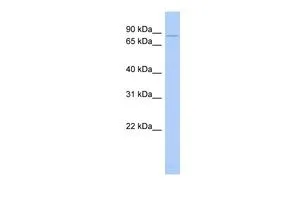
Figure 1. Western blot analysis of CHM using anti-CHM antibody (A00814-1). Electrophoresis was performed on a 5-20% SDS-PAGE gel at 70V (Stacking gel) / 90V (Resolving gel) for 2-3 hours. The sample well of each lane was loaded with 30 ug of sample under reducing conditions. Lane 1: human Hela whole cell lysates, Lane 2: human A431 whole cell lysates, Lane 3: human 293T whole cell lysates, Lane 4: rat liver tissue lysates, Lane 5: mouse liver tissue lysates. After electrophoresis, proteins were transferred to a nitrocellulose membrane at 150 mA for 50-90 minutes. Blocked the membrane with 5% non-fat milk/TBS for 1.5 hour at RT. The membrane was incubated with rabbit anti-CHM antigen affinity purified polyclonal antibody (Catalog # A00814-1) at 0.5 microg/mL overnight at 4°C, then washed with TBS-0.1%Tween 3 times with 5 minutes each and probed with a goat anti-rabbit IgG-HRP secondary antibody at a dilution of 1:5000 for 1.5 hour at RT. The signal is developed using an Enhanced Chemiluminescent detection (ECL) kit (Catalog # EK1002) with Tanon 5200 system. A specific band was detected for CHM at approximately 73 kDa. The expected band size for CHM is at 100 kDa.
Figure 1. Western blot analysis of CHM using anti-CHM antibody (A00814-1). Electrophoresis was performed on a 5-20% SDS-PAGE gel at 70V (Stacking gel) / 90V (Resolving gel) for 2-3 hours. The sample well of each lane was loaded with 30 ug of sample under reducing conditions. Lane 1: human Hela whole cell lysates, Lane 2: human A431 whole cell lysates, Lane 3: human 293T whole cell lysates, Lane 4: rat liver tissue lysates, Lane 5: mouse liver tissue lysates. After electrophoresis, proteins were transferred to a nitrocellulose membrane at 150 mA for 50-90 minutes. Blocked the membrane with 5% non-fat milk/TBS for 1.5 hour at RT. The membrane was incubated with rabbit anti-CHM antigen affinity purified polyclonal antibody (Catalog # A00814-1) at 0.5 microg/mL overnight at 4°C, then washed with TBS-0.1%Tween 3 times with 5 minutes each and probed with a goat anti-rabbit IgG-HRP secondary antibody at a dilution of 1:5000 for 1.5 hour at RT. The signal is developed using an Enhanced Chemiluminescent detection (ECL) kit (Catalog # EK1002) with Tanon 5200 system. A specific band was detected for CHM at approximately 73 kDa. The expected band size for CHM is at 100 kDa.
Anti-CHM Antibody Picoband(r)
A00814-1-CARRIER-FREE
ApplicationsWestern Blot, ELISA
Product group Antibodies
ReactivityHuman, Mouse, Rat
TargetCHM
Overview
- SupplierBoster Bio
- Product NameAnti-CHM Antibody Picoband(r)
- Delivery Days Customer9
- ApplicationsWestern Blot, ELISA
- CertificationResearch Use Only
- ClonalityPolyclonal
- Concentration500 ug/ml
- Gene ID1121
- Target nameCHM
- Target descriptionCHM Rab escort protein
- Target synonymsDXS540, GGTA, HSD-32, REP-1, TCD, rab proteins geranylgeranyltransferase component A 1, CHM, Rab escort protein 1, choroideremia (Rab escort protein 1)
- HostRabbit
- IsotypeIgG
- Protein IDP24386
- Protein NameRab proteins geranylgeranyltransferase component A 1
- Scientific DescriptionBoster Bio Anti-CHM Antibody Picoband® catalog # A00814-1. Tested in ELISA, WB applications. This antibody reacts with Human, Mouse, Rat. The brand Picoband indicates this is a premium antibody that guarantees superior quality, high affinity, and strong signals with minimal background in Western blot applications. Only our best-performing antibodies are designated as Picoband, ensuring unmatched performance.
- ReactivityHuman, Mouse, Rat
- Storage Instruction-20°C,2°C to 8°C
- UNSPSC12352203
Related products
Product group Antibodies
CHM AntibodyCSB-PA005356LA01HU
ApplicationsImmunoFluorescence, ELISA, ImmunoHistoChemistry
ReactivityHuman
TargetCHM
- SizePrice

Product group Antibodies
Anti-CHM AntibodyHPA003231
ApplicationsImmunoHistoChemistry
ReactivityHuman
TargetCHM
- SizePrice
Product group Antibodies

CHM / REP1 AntibodyLS-C483002
ApplicationsWestern Blot
ReactivityHuman
TargetCHM
- SizePrice
Product group Antibodies
Chm Polyclonal AntibodyCAC08312
ApplicationsImmunoFluorescence, ELISA, ImmunoHistoChemistry
TargetCHM
- SizePrice
Product group Antibodies
Choroideremia antibody, N-termGTX48937
ApplicationsWestern Blot
ReactivityHuman
TargetCHM
- SizePrice
Product group Antibodies
Anti-CHM Antibody144-08345
ApplicationsWestern Blot
ReactivityHuman
TargetCHM
- SizePrice
Product group Antibodies
CHM Recombinant AntibodyBSM-62531R
ApplicationsImmunoFluorescence, Western Blot, ImmunoCytoChemistry
ReactivityHuman, Mouse, Rat
TargetCHM
- SizePrice