


Western blot analysis of NLRP3 using anti-NLRP3 antibody (PA1665).
Electrophoresis was performed on a 8% SDS-PAGE gel at 80V (Stacking gel) / 120V (Resolving gel) for 2 hours. The sample well of each lane was loaded with 30 ug of sample under reducing conditions.
Lane 1: rat thymus tissue lysates,
Lane 2: mouse thyus tissue lysates,
Lane 3: mouse RAW264.7 whole cell lysates,
Lane 4: mouse J774A.1 whole cell lysates.
After electrophoresis, proteins were transferred to a nitrocellulose membrane at 150 mA for 50-90 minutes. Blocked the membrane with 5% non-fat milk/TBS for 1.5 hour at RT. The membrane was incubated with rabbit anti-NLRP3 antigen affinity purified polyclonal antibody (PA1665) at 0.5 μg/mL overnight at 4°C, then washed with TBS-0.1%Tween 3 times with 5 minutes each and probed with a goat anti-rabbit IgG-HRP secondary antibody (Catalog # BA1054) at a dilution of 1:5000 for 1.5 hour at RT. The signal is developed using an ECL Plus Western Blotting Substrate (Catalog # AR1196-200) with Tanon 5200 system. A specific band was detected for NLRP3 at approximately 110 kDa. The expected band size for NLRP3 is at 118kDa.
.
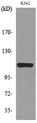
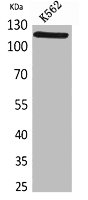
Electrophoresis was performed on a 8% SDS-PAGE gel at 80V (Stacking gel) / 120V (Resolving gel) for 2 hours. The sample well of each lane was loaded with 30 ug of sample under reducing conditions.
Lane 1:human THP-1 whole cell lysates.
After electrophoresis, proteins were transferred to a nitrocellulose membrane at 150 mA for 50-90 minutes. Blocked the membrane with 5% non-fat milk/TBS for 1.5 hour at RT. The membrane was incubated with rabbit anti-NLRP3 antigen affinity purified polyclonal antibody (PA1665) at 0.5 μg/mL overnight at 4°C, then washed with TBS-0.1%Tween 3 times with 5 minutes each and probed with a goat anti-rabbit IgG-HRP secondary antibody (Catalog # BA1054) at a dilution of 1:5000 for 1.5 hour at RT. The signal is developed using an ECL Plus Western Blotting Substrate (Catalog # AR1196-200) with Tanon 5200 system. A specific band was detected for NLRP3 at approximately 110 kDa. The expected band size for NLRP3 is at 118kDa.")
.
NLRP3 was detected in a paraffin-embedded section of human colon cancer tissue. Heat mediated antigen retrieval was performed in EDTA buffer (pH 8.0, epitope retrieval solution). The tissue section was blocked with 10% goat serum. The tissue section was then incubated with 2 μg/ml rabbit anti-NLRP3 Antibody (PA1665) overnight at 4°C. Peroxidase Conjugated Goat Anti-rabbit IgG was used as secondary antibody and incubated for 30 minutes at 37°C. The tissue section was developed using HRP Conjugated Rabbit IgG Super Vision Assay Kit (Catalog # SV0002) with DAB as the chromogen.")
.
Overlay histogram showing THP-1 cells stained with PA1665 (Blue line). To facilitate intracellular staining, cells were fixed with 4% paraformaldehyde and permeabilized with permeabilization buffer. The cells were blocked with 10% normal goat serum. And then incubated with rabbit anti-NLRP3 Antibody (PA1665, 1 μg/1x106 cells) for 30 min at 20°C. DyLight®488 conjugated goat anti-rabbit IgG (BA1127, 5-10 μg/1x106 cells) was used as secondary antibody for 30 minutes at 20°C. Isotype control antibody (Green line) was rabbit IgG (1 μg/1x106) used under the same conditions. Unlabelled sample without incubation with primary antibody and secondary antibody (Red line) was used as a blank control.")
Western blot analysis of NLRP3 using anti-NLRP3 antibody (PA1665).
Electrophoresis was performed on a 8% SDS-PAGE gel at 80V (Stacking gel) / 120V (Resolving gel) for 2 hours. The sample well of each lane was loaded with 30 ug of sample under reducing conditions.
Lane 1: rat thymus tissue lysates,
Lane 2: mouse thyus tissue lysates,
Lane 3: mouse RAW264.7 whole cell lysates,
Lane 4: mouse J774A.1 whole cell lysates.
After electrophoresis, proteins were transferred to a nitrocellulose membrane at 150 mA for 50-90 minutes. Blocked the membrane with 5% non-fat milk/TBS for 1.5 hour at RT. The membrane was incubated with rabbit anti-NLRP3 antigen affinity purified polyclonal antibody (PA1665) at 0.5 μg/mL overnight at 4°C, then washed with TBS-0.1%Tween 3 times with 5 minutes each and probed with a goat anti-rabbit IgG-HRP secondary antibody (Catalog # BA1054) at a dilution of 1:5000 for 1.5 hour at RT. The signal is developed using an ECL Plus Western Blotting Substrate (Catalog # AR1196-200) with Tanon 5200 system. A specific band was detected for NLRP3 at approximately 110 kDa. The expected band size for NLRP3 is at 118kDa.
Anti-CIAS1/NALP3/NLRP3 Antibody Picoband(r)
PA1665
ApplicationsFlow Cytometry, Western Blot, ImmunoHistoChemistry
Product group Antibodies
ReactivityHamster, Human, Mouse, Rat
TargetNLRP3
Overview
- SupplierBoster Bio
- Product NameAnti-CIAS1/NALP3/NLRP3 Antibody Picoband(r)
- Delivery Days Customer9
- Application Supplier NoteTested Species: In-house tested species with positive results. Predicted Species: Species predicted to be fit for the product based on sequence similarities. By Heat: Boiling the paraffin sections in 10mM citrate buffer, pH6.0, for 20mins is required for the staining of formalin/paraffin sections. Other applications have not been tested. Optimal dilutions should be determined by end users.
- ApplicationsFlow Cytometry, Western Blot, ImmunoHistoChemistry
- Applications SupplierIHP, WB, IHC
- CertificationResearch Use Only
- ClonalityPolyclonal
- Concentration500 ug/ml
- Gene ID114548
- Target nameNLRP3
- Target descriptionNLR family pyrin domain containing 3
- Target synonymsAGTAVPRL, AII, AVP, C1orf7, CIAS1, CLR1.1, DFNA34, FCAS, FCAS1, FCU, KEFH, MWS, NALP3, PYPAF1, NACHT, LRR and PYD domains-containing protein 3, NACHT domain-, leucine-rich repeat-, and PYD-containing protein 3, NACHT, LRR and PYD containing protein 3, PYRIN-containing APAF1-like protein 1, caterpiller protein 1.1, cold autoinflammatory syndrome 1 protein, cold-induced autoinflammatory syndrome 1 protein, cryopyrin, cryopyrin, NACHT, LRR and PYD domains - containing protein 3, deafness, autosomal dominant 34, nucleotide-binding oligomerization domain, leucine rich repeat and pyrin domain containing 3
- HostRabbit
- IsotypeIgG
- Protein IDQ96P20
- Protein NameNACHT, LRR and PYD domains-containing protein 3
- Scientific DescriptionBoster Bio Anti-CIAS1/NALP3/NLRP3 Antibody catalog # PA1665. Tested in Flow Cytometry, IHC, WB applications. This antibody reacts with Human, Mouse, Rat. The brand Picoband indicates this is a premium antibody that guarantees superior quality, high affinity, and strong signals with minimal background in Western blot applications. Only our best-performing antibodies are designated as Picoband, ensuring unmatched performance.
- ReactivityHamster, Human, Mouse, Rat
- Reactivity SupplierHuman, Mouse, Rat, Hamster
- Storage Instruction-20°C,2°C to 8°C
- UNSPSC12352203
References
- Yang L, Xing W, Shi Y, et al. Stress-induced NLRP3 inflammasome activation and myelin alterations in the hippocampus of PTSD rats. Neuroscience. 2024,555:156-166. doi: 10.1016/j.neuroscience.2024.07.028Read this paper
- Chen S, Zhu L, Fang X, et al. Alloferon Mitigates LPS-Induced Endometritis by Attenuating the NLRP3/CASP1/IL-1β/IL-18 Signaling Cascade. Inflammation. 2025,48(2):730-746. doi: 10.1007/s10753-024-02083-6Read this paper
- Chi Q, Xia Y, Luo D, et al. In vitro and in silico analyses reveal the toxicity of metolachlor to grass carp hepatocytes and the antagonism of melatonin. Pestic Biochem Physiol. 2024,202:105930. doi: 10.1016/j.pestbp.2024.105930Read this paper
- Chen J, Hu J, Guo X, et al. Apolipoprotein O modulates cholesterol metabolism via NRF2/CYB5R3 independent of LDL receptor. Cell Death Dis. 2024,15(6):389. doi: 10.1038/s41419-024-06778-4Read this paper
- Jia R, Ma H, Hao H, et al. Metformin inhibits activation of NLRP3 inflammasome and inflammatory response in preeclamptic rats. Gene. 2024,919:148509. doi: 10.1016/j.gene.2024.148509Read this paper
- Zhang R, Zhu Z, Ma Y, et al. Rhizoma Alismatis Decoction improved mitochondrial dysfunction to alleviate SASP by enhancing autophagy flux and apoptosis in hyperlipidemia acute pancreatitis. Phytomedicine. 2024,129:155629. doi: 10.1016/j.phymed.2024.155629Read this paper
- Shen J, Xu J, Wen Y, et al. Carnosine ameliorates postoperative cognitive dysfunction of aged rats by limiting astrocytes pyroptosis. Neurotherapeutics. 2024,21(4):e00359. doi: 10.1016/j.neurot.2024.e00359Read this paper
- Li J, Teng D, Jia W, et al. PLD2 deletion ameliorates sepsis-induced cardiomyopathy by suppressing cardiomyocyte pyroptosis via the NLRP3/caspase 1/GSDMD pathway. Inflamm Res. 2024,73(6):1033-1046. doi: 10.1007/s00011-024-01881-wRead this paper
- Li X, Cheng L, Liu X, et al. Dopamine promotes Klebsiella quasivariicola proliferation and inflammatory response in the presence of macrophages. Front Cell Infect Microbiol. 2024,14:1322113. doi: 10.3389/fcimb.2024.1322113Read this paper
- Chai Y, Gu X, Zhang H, et al. Phoenixin 20 ameliorates pulmonary arterial hypertension via inhibiting inflammation and oxidative stress. Aging (Albany NY). 2024,16(6):5027-5037. doi: 10.18632/aging.205468Read this paper
Datasheet
MSDS
Related products
Product group Antibodies
Anti-NLRP3 AntibodyA100943
ApplicationsWestern Blot, ELISA
ReactivityHuman
- SizePrice
Product group Antibodies
Anti-NLRP3 Antibody144-05652
ApplicationsImmunoFluorescence, Western Blot
ReactivityHuman, Mouse, Rat
TargetNLRP3
- SizePrice

Product group Antibodies
Anti-NLRP3 AntibodyAMAB90569
ApplicationsWestern Blot, ImmunoCytoChemistry
ReactivityHuman
TargetNLRP3
- SizePrice

Product group Antibodies
Anti-CIAS1/NALP3/NLRP3 Antibody Picoband(r)A00034-2-CARRIER-FREE
ApplicationsFlow Cytometry, Western Blot, ELISA
ReactivityHuman, Mouse, Rat
TargetNLRP3
- SizePrice

Product group Antibodies
References
NALP3/CIAS1 Polyclonal AntibodyBS-10021R
ApplicationsImmunoFluorescence, Western Blot, ImmunoHistoChemistry, ImmunoHistoChemistry Frozen, ImmunoHistoChemistry Paraffin
ReactivityCanine, Human, Mouse, Rat
TargetNLRP3
- SizePrice
Product group Antibodies

ApplicationsELISA
ReactivityBovine, Canine, Human, Mouse
TargetNLRP3
- SizePrice
Product group Antibodies
NLRP3 AntibodyCSB-PA005618
ApplicationsWestern Blot, ELISA
ReactivityHuman
TargetNLRP3
- SizePrice
Product group Antibodies
ApplicationsImmunoPrecipitation, Western Blot, ImmunoCytoChemistry, ImmunoHistoChemistry
TargetNLRP3
- SizePrice
Product group Antibodies

NALP3 / NLRP3 Antibody (Internal)LS-C368954
ApplicationsWestern Blot
ReactivityHuman
TargetNLRP3
- SizePrice
![Boiled and unboiled THP-1 whole cell and membrane extracts (30 μg) were separated by 7.5% SDS-PAGE, and the membrane was blotted with NLRP3 antibody [C3], C-term (GTX106313) diluted at 1:1000. The HRP-conjugated anti-rabbit IgG antibody (GTX213110-01) was used to detect the primary antibody. (WCE: whole cell extract; ME: membrane extract)](https://www.genetex.com/upload/website/prouct_img/normal/GTX106313/GTX106313_44454_20211008_WB_Fraction_w_23060120_958.webp)
Product group Antibodies
NLRP3 antibody [C3], C-termGTX106313
ApplicationsImmunoPrecipitation, Western Blot
ReactivityHuman
TargetNLRP3
- SizePrice