


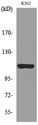
Figure 1. Western blot analysis of NLRP3 using anti-NLRP3 antibody (A00034-2). Electrophoresis was performed on a 5-20% SDS-PAGE gel at 70V (Stacking gel) / 90V (Resolving gel) for 2-3 hours. The sample well of each lane was loaded with 30ug of sample under reducing conditions. Lane 1: human Hela whole cell lysates, Lane 2: human U-87MG whole cell lysates, Lane 3: rat spleen tissue lysates, Lane 4: rat PC-12 whole cell lysates, Lane 5: mouse thymus tissue lysates, Lane 6: mouse lung tissue lysates, Lane 7: mouse spleen tissue lysates. After Electrophoresis, proteins were transferred to a Nitrocellulose membrane at 150mA for 50-90 minutes. Blocked the membrane with 5% Non-fat Milk/ TBS for 1.5 hour at RT. The membrane was incubated with rabbit anti-NLRP3 antigen affinity purified polyclonal antibody (Catalog # A00034-2) at 0.5 microg/mL overnight at 4°C, then washed with TBS-0.1%Tween 3 times with 5 minutes each and probed with a goat anti-rabbit IgG-HRP secondary antibody at a dilution of 1:5000 for 1.5 hour at RT. The signal is developed using an Enhanced Chemiluminescent detection (ECL) kit (Catalog # EK1002) with Tanon 5200 system. A specific band was detected for NLRP3 at approximately 118KD. The expected band size for NLRP3 is at 118KD.
. Overlay histogram showing THP-1 cells stained with A00034-2 (Blue line).To facilitate intracellular staining, cells were fixed with 4% paraformaldehyde and permeabilized with permeabilization buffer. The cells were blocked with 10% normal goat serum. And then incubated with rabbit anti-NLRP3 Antibody (A00034-2, 1microg/1x106 cells) for 30 min at 20°C. DyLight®488 conjugated goat anti-rabbit IgG (BA1127, 5-10microg/1x106 cells) was used as secondary antibody for 30 minutes at 20°C. Isotype control antibody (Green line) was rabbit IgG (1microg/1x106) used under the same conditions. Unlabelled sample without incubation with primary antibody and secondary antibody (Red line) was used as a blank control.")
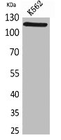
Figure 1. Western blot analysis of NLRP3 using anti-NLRP3 antibody (A00034-2). Electrophoresis was performed on a 5-20% SDS-PAGE gel at 70V (Stacking gel) / 90V (Resolving gel) for 2-3 hours. The sample well of each lane was loaded with 30ug of sample under reducing conditions. Lane 1: human Hela whole cell lysates, Lane 2: human U-87MG whole cell lysates, Lane 3: rat spleen tissue lysates, Lane 4: rat PC-12 whole cell lysates, Lane 5: mouse thymus tissue lysates, Lane 6: mouse lung tissue lysates, Lane 7: mouse spleen tissue lysates. After Electrophoresis, proteins were transferred to a Nitrocellulose membrane at 150mA for 50-90 minutes. Blocked the membrane with 5% Non-fat Milk/ TBS for 1.5 hour at RT. The membrane was incubated with rabbit anti-NLRP3 antigen affinity purified polyclonal antibody (Catalog # A00034-2) at 0.5 microg/mL overnight at 4°C, then washed with TBS-0.1%Tween 3 times with 5 minutes each and probed with a goat anti-rabbit IgG-HRP secondary antibody at a dilution of 1:5000 for 1.5 hour at RT. The signal is developed using an Enhanced Chemiluminescent detection (ECL) kit (Catalog # EK1002) with Tanon 5200 system. A specific band was detected for NLRP3 at approximately 118KD. The expected band size for NLRP3 is at 118KD.
Anti-CIAS1/NALP3/NLRP3 Antibody Picoband(r)
A00034-2-CARRIER-FREE
ApplicationsFlow Cytometry, Western Blot, ELISA
Product group Antibodies
ReactivityHuman, Mouse, Rat
TargetNLRP3
Overview
- SupplierBoster Bio
- Product NameAnti-CIAS1/NALP3/NLRP3 Antibody Picoband(r)
- Delivery Days Customer9
- ApplicationsFlow Cytometry, Western Blot, ELISA
- CertificationResearch Use Only
- ClonalityPolyclonal
- Concentration500 ug/ml
- Gene ID114548
- Target nameNLRP3
- Target descriptionNLR family pyrin domain containing 3
- Target synonymsAGTAVPRL, AII, AVP, C1orf7, CIAS1, CLR1.1, DFNA34, FCAS, FCAS1, FCU, KEFH, MWS, NALP3, PYPAF1, NACHT, LRR and PYD domains-containing protein 3, NACHT domain-, leucine-rich repeat-, and PYD-containing protein 3, NACHT, LRR and PYD containing protein 3, PYRIN-containing APAF1-like protein 1, caterpiller protein 1.1, cold autoinflammatory syndrome 1 protein, cold-induced autoinflammatory syndrome 1 protein, cryopyrin, cryopyrin, NACHT, LRR and PYD domains - containing protein 3, deafness, autosomal dominant 34, nucleotide-binding oligomerization domain, leucine rich repeat and pyrin domain containing 3
- HostRabbit
- IsotypeIgG
- Protein IDQ96P20
- Protein NameNACHT, LRR and PYD domains-containing protein 3
- Scientific DescriptionBoster Bio Anti-CIAS1/NALP3/NLRP3 Antibody Picoband® catalog # A00034-2. Tested in ELISA, Flow Cytometry, WB applications. This antibody reacts with Human, Mouse, Rat. The brand Picoband indicates this is a premium antibody that guarantees superior quality, high affinity, and strong signals with minimal background in Western blot applications. Only our best-performing antibodies are designated as Picoband, ensuring unmatched performance.
- ReactivityHuman, Mouse, Rat
- Storage Instruction-20°C,2°C to 8°C
- UNSPSC12352203
Related products
Product group Antibodies
Anti-NLRP3 AntibodyA100943
ApplicationsWestern Blot, ELISA
ReactivityHuman
- SizePrice
Product group Antibodies
Anti-NLRP3 Antibody144-05652
ApplicationsImmunoFluorescence, Western Blot
ReactivityHuman, Mouse, Rat
TargetNLRP3
- SizePrice

Product group Antibodies
Anti-NLRP3 AntibodyAMAB90569
ApplicationsWestern Blot, ImmunoCytoChemistry
ReactivityHuman
TargetNLRP3
- SizePrice

Product group Antibodies
References
NALP3/CIAS1 Polyclonal AntibodyBS-10021R
ApplicationsImmunoFluorescence, Western Blot, ImmunoHistoChemistry, ImmunoHistoChemistry Frozen, ImmunoHistoChemistry Paraffin
ReactivityCanine, Human, Mouse, Rat
TargetNLRP3
- SizePrice
Product group Antibodies

ApplicationsELISA
ReactivityBovine, Canine, Human, Mouse
TargetNLRP3
- SizePrice
Product group Antibodies
NLRP3 AntibodyCSB-PA005618
ApplicationsWestern Blot, ELISA
ReactivityHuman
TargetNLRP3
- SizePrice
Product group Antibodies
ApplicationsImmunoPrecipitation, Western Blot, ImmunoCytoChemistry, ImmunoHistoChemistry
TargetNLRP3
- SizePrice
Product group Antibodies

NALP3 / NLRP3 Antibody (Internal)LS-C368954
ApplicationsWestern Blot
ReactivityHuman
TargetNLRP3
- SizePrice
![Boiled and unboiled THP-1 whole cell and membrane extracts (30 μg) were separated by 7.5% SDS-PAGE, and the membrane was blotted with NLRP3 antibody [C3], C-term (GTX106313) diluted at 1:1000. The HRP-conjugated anti-rabbit IgG antibody (GTX213110-01) was used to detect the primary antibody. (WCE: whole cell extract; ME: membrane extract)](https://www.genetex.com/upload/website/prouct_img/normal/GTX106313/GTX106313_44454_20211008_WB_Fraction_w_23060120_958.webp)
Product group Antibodies
NLRP3 antibody [C3], C-termGTX106313
ApplicationsImmunoPrecipitation, Western Blot
ReactivityHuman
TargetNLRP3
- SizePrice