


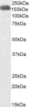
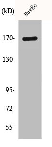
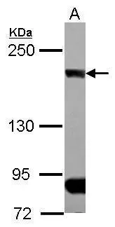
Figure 1. Western blot analysis of CLIP170 using anti-CLIP170 antibody (M08907). Electrophoresis was performed on a 5-20% SDS-PAGE gel at 70V (Stacking gel) / 90V (Resolving gel) for 2-3 hours. The sample well of each lane was loaded with 30 ug of sample under reducing conditions. Lane 1: human Hela whole cell lysates, Lane 2: human SiHa whole cell lysates, Lane 3: human A549 whoel cell lysates, Lane 4: human MCF-7 whole cell lysates, Lane 5: rat brain tissue lysates, Lane 6: mouse brain tissue lysates. After electrophoresis, proteins were transferred to a nitrocellulose membrane at 150 mA for 50-90 minutes. Blocked the membrane with 5% non-fat milk/TBS for 1.5 hour at RT. The membrane was incubated with rabbit anti-CLIP170 antigen affinity purified monoclonal antibody (M08907) at 1:500 overnight at 4°C, then washed with TBS-0.1%Tween 3 times with 5 minutes each and probed with a goat anti-rabbit IgG-HRP secondary antibody at a dilution of 1:500 for 1.5 hour at RT. The signal is developed using an Enhanced Chemiluminescent detection (ECL) kit (Catalog # EK1002) with Tanon 5200 system. A specific band was detected for CLIP170 at approximately 170 kDa. The expected band size for CLIP170 is at 162 kDa.
Figure 1. Western blot analysis of CLIP170 using anti-CLIP170 antibody (M08907). Electrophoresis was performed on a 5-20% SDS-PAGE gel at 70V (Stacking gel) / 90V (Resolving gel) for 2-3 hours. The sample well of each lane was loaded with 30 ug of sample under reducing conditions. Lane 1: human Hela whole cell lysates, Lane 2: human SiHa whole cell lysates, Lane 3: human A549 whoel cell lysates, Lane 4: human MCF-7 whole cell lysates, Lane 5: rat brain tissue lysates, Lane 6: mouse brain tissue lysates. After electrophoresis, proteins were transferred to a nitrocellulose membrane at 150 mA for 50-90 minutes. Blocked the membrane with 5% non-fat milk/TBS for 1.5 hour at RT. The membrane was incubated with rabbit anti-CLIP170 antigen affinity purified monoclonal antibody (M08907) at 1:500 overnight at 4°C, then washed with TBS-0.1%Tween 3 times with 5 minutes each and probed with a goat anti-rabbit IgG-HRP secondary antibody at a dilution of 1:500 for 1.5 hour at RT. The signal is developed using an Enhanced Chemiluminescent detection (ECL) kit (Catalog # EK1002) with Tanon 5200 system. A specific band was detected for CLIP170 at approximately 170 kDa. The expected band size for CLIP170 is at 162 kDa.
Anti-CLIP170 Rabbit Monoclonal Antibody
M08907
ApplicationsWestern Blot, ImmunoHistoChemistry
Product group Antibodies
ReactivityHuman, Mouse, Rat
TargetCLIP1
Overview
- SupplierBoster Bio
- Product NameAnti-CLIP170 Rabbit Monoclonal Antibody
- Delivery Days Customer9
- ApplicationsWestern Blot, ImmunoHistoChemistry
- CertificationResearch Use Only
- ClonalityMonoclonal
- Clone ID29C06
- Gene ID6249
- Target nameCLIP1
- Target descriptionCAP-Gly domain containing linker protein 1
- Target synonymsCLIP, CLIP-170, CLIP170, CYLN1, RSN, CAP-Gly domain-containing linker protein 1, cytoplasmic linker protein 1, cytoplasmic linker protein 170 alpha-2, cytoplasmic linker protein CLIP-170, restin (Reed-Steinberg cell-expressed intermediate filament-associated protein)
- HostRabbit
- IsotypeIgG
- Protein IDP30622
- Protein NameCAP-Gly domain-containing linker protein 1
- Scientific DescriptionBoster Bio Anti-CLIP170 Rabbit Monoclonal Antibody catalog # M08907. Tested in WB, IHC applications. This antibody reacts with Human, Mouse, Rat.
- ReactivityHuman, Mouse, Rat
- Storage Instruction-20°C
- UNSPSC12352203
Related products
Product group Antibodies
ApplicationsWestern Blot, ELISA
ReactivityHuman
- SizePrice
Product group Antibodies
Anti-CLIP1 Antibody144-07722
ApplicationsWestern Blot, ImmunoHistoChemistry
ReactivityHuman, Mouse, Rat
TargetCLIP1
- SizePrice

Product group Antibodies
Anti-CLIP1 AntibodyAMAB91321
ApplicationsWestern Blot, ImmunoCytoChemistry, ImmunoHistoChemistry
ReactivityHuman
TargetCLIP1
- SizePrice
Product group Antibodies

CLIP1 / CLIP-170 AntibodyLS-C747597
ApplicationsWestern Blot
ReactivityHuman, Mouse, Rat
TargetCLIP1
- SizePrice
Product group Antibodies
ApplicationsFlow Cytometry, Western Blot, ImmunoCytoChemistry
ReactivityHuman, Mouse, Rat
TargetCLIP1
- SizePrice
Product group Antibodies
CLIP1 AntibodyCSB-PA001702
ApplicationsWestern Blot, ELISA
ReactivityHuman, Mouse, Rat
TargetCLIP1
- SizePrice
Product group Antibodies

ApplicationsWestern Blot, ELISA
ReactivityCanine, Human, Mouse, Porcine, Rat
TargetCLIP1
- SizePrice
Product group Antibodies
CLIP170 antibody [N1], N-termGTX117504
ApplicationsImmunoFluorescence, Western Blot, ImmunoCytoChemistry
ReactivityHuman, Mouse, Rat
TargetCLIP1
- SizePrice
Product group Antibodies
Anti-CLIP1 AntibodyCAB7722
ApplicationsWestern Blot, ELISA
ReactivityHuman
TargetCLIP1
- SizePrice