


Immunofluorescence staining of mouse brain cortex shows strong cytoplasmic positivity in neuropil.
Immunofluorescence staining of mouse brain cortex shows strong cytoplasmic positivity in neuropil.
Anti-CNTN1 Antibody
HPA070467
ApplicationsImmunoHistoChemistry
Product group Antibodies
ReactivityHuman, Mouse
TargetCNTN1
Overview
- SupplierAtlas Antibodies
- Product NameAnti-CNTN1 Antibody
- Delivery Days Customer4
- ApplicationsImmunoHistoChemistry
- CertificationResearch Use Only
- ClonalityPolyclonal
- ConjugateUnconjugated
- Gene ID1272
- Target nameCNTN1
- Target descriptioncontactin 1
- Target synonymsCMYO12, CMYP12, F3, GP135, MYPCN, contactin-1, glycoprotein gP135, neural cell surface protein F3
- HostRabbit
- IsotypeIgG
- Protein IDQ12860
- Protein NameContactin-1
- Scientific DescriptionRecombinant Protein Epitope Signature Tag (PrEST) antigen sequence
- ReactivityHuman, Mouse
- Storage Instruction-20°C,2°C to 8°C
- UNSPSC41116161
Datasheet
MSDS
Related products
Product group Antibodies
CNTN1 AntibodyCSB-PA004825
ApplicationsWestern Blot, ELISA
ReactivityHuman, Mouse, Rat
TargetCNTN1
- SizePrice
Product group Antibodies
Anti-Contactin 1 AntibodyA306547
ApplicationsWestern Blot, ImmunoHistoChemistry
ReactivityHuman, Mouse, Rat
- SizePrice
Product group Antibodies

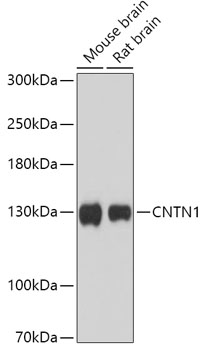
CNTN1 / gp135 / Contactin 1 AntibodyLS-C831132
ApplicationsWestern Blot, ELISA, ImmunoHistoChemistry
ReactivityHuman, Mouse, Rat
TargetCNTN1
- SizePrice
Product group Antibodies

Goat anti-contactin 1EB12273
ApplicationsWestern Blot, ELISA, ImmunoHistoChemistry
ReactivityHuman, Mouse, Porcine, Rat
TargetCNTN1
- SizePrice

Product group Antibodies
Anti-Contactin 1/CNTN1 Antibody Picoband(r)A04538-2-CARRIER-FREE
ApplicationsImmunoFluorescence, Western Blot, ELISA, ImmunoCytoChemistry
ReactivityHuman, Mouse, Rat
TargetCNTN1
- SizePrice
Product group Antibodies
Cntn1 Polyclonal AntibodyCAC11289
ApplicationsImmunoFluorescence, ELISA
TargetCNTN1
- SizePrice
![Contactin 1 antibody detects Contactin 1 protein by immunohistochemical analysis. Sample: Frozen sectioned adult mouse retina. Green: Contactin 1 protein stained by Contactin 1 antibody (GTX101735) diluted at 1:250. Red: beta Tubulin 3/ TUJ1, stained by beta Tubulin 3/ TUJ1 antibody [GT11710] (GTX631836) diluted at 1:250. Blue: Fluoroshield with DAPI (GTX30920).](https://www.genetex.com/upload/website/prouct_img/normal/GTX101735/GTX101735_40128_20160830_IHC-Fr_w_23060100_879.webp)
Product group Antibodies
Contactin 1 antibodyGTX101735
ApplicationsImmunoHistoChemistry, ImmunoHistoChemistry Frozen
ReactivityHuman, Mouse
TargetCNTN1
- SizePrice
Product group Antibodies
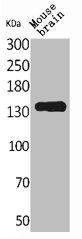
Anti-CNTN1 Antibody144-64936
ApplicationsWestern Blot
ReactivityHuman, Mouse, Rat
TargetCNTN1
- SizePrice