


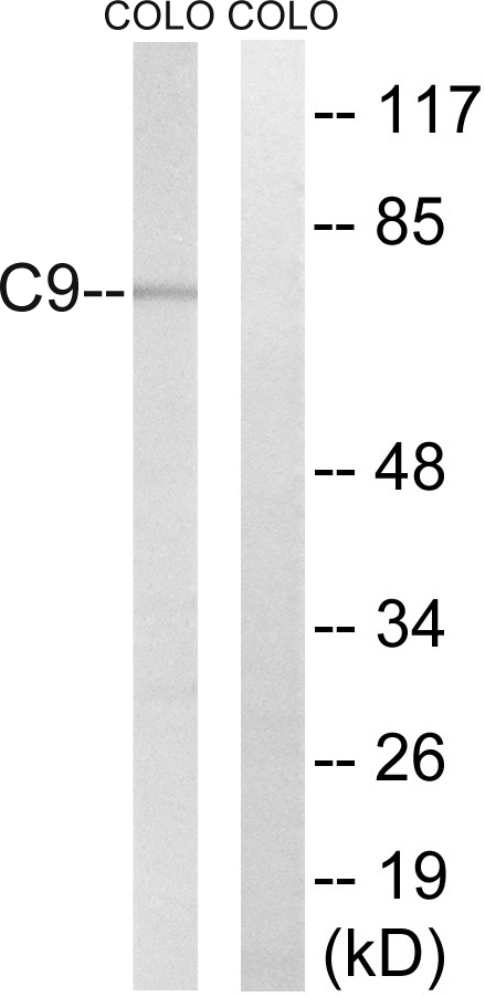
Figure 1. Western blot analysis of Complement C9 using anti-Complement C9 antibody (A01010-2). Electrophoresis was performed on a 5-20% SDS-PAGE gel at 70V (Stacking gel) / 90V (Resolving gel) for 2-3 hours. The sample well of each lane was loaded with 50ug of sample under reducing conditions. Lane 1: human placenta tissue lysates. After Electrophoresis, proteins were transferred to a Nitrocellulose membrane at 150mA for 50-90 minutes. Blocked the membrane with 5% Non-fat Milk/ TBS for 1.5 hour at RT. The membrane was incubated with rabbit anti-Complement C9 antigen affinity purified polyclonal antibody (Catalog # A01010-2) at 0.5 microg/mL overnight at 4°C, then washed with TBS-0.1%Tween 3 times with 5 minutes each and probed with a goat anti-rabbit IgG-HRP secondary antibody at a dilution of 1:10000 for 1.5 hour at RT. The signal is developed using an Enhanced Chemiluminescent detection (ECL) kit (Catalog # EK1002) with Tanon 5200 system. A specific band was detected for Complement C9 at approximately 80KD. The expected band size for Complement C9 is at 63KD.
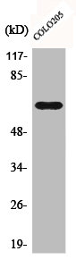
. Electrophoresis was performed on a 5-20% SDS-PAGE gel at 70V (Stacking gel) / 90V (Resolving gel) for 2-3 hours. The sample well of each lane was loaded with 50ug of sample under reducing conditions. Lane 1: mouse liver tissue lysates, Lane 2: mouse lung tissue lysates, After Electrophoresis, proteins were transferred to a Nitrocellulose membrane at 150mA for 50-90 minutes. Blocked the membrane with 5% Non-fat Milk/ TBS for 1.5 hour at RT. The membrane was incubated with rabbit anti-Complement C9 antigen affinity purified polyclonal antibody (Catalog # A01010-2) at 0.5 microg/mL overnight at 4°C, then washed with TBS-0.1%Tween 3 times with 5 minutes each and probed with a goat anti-rabbit IgG-HRP secondary antibody at a dilution of 1:10000 for 1.5 hour at RT. The signal is developed using an Enhanced Chemiluminescent detection (ECL) kit (Catalog # EK1002) with Tanon 5200 system. A specific band was detected for Complement C9 at approximately 80KD. The expected band size for Complement C9 is at 63KD.")
Figure 1. Western blot analysis of Complement C9 using anti-Complement C9 antibody (A01010-2). Electrophoresis was performed on a 5-20% SDS-PAGE gel at 70V (Stacking gel) / 90V (Resolving gel) for 2-3 hours. The sample well of each lane was loaded with 50ug of sample under reducing conditions. Lane 1: human placenta tissue lysates. After Electrophoresis, proteins were transferred to a Nitrocellulose membrane at 150mA for 50-90 minutes. Blocked the membrane with 5% Non-fat Milk/ TBS for 1.5 hour at RT. The membrane was incubated with rabbit anti-Complement C9 antigen affinity purified polyclonal antibody (Catalog # A01010-2) at 0.5 microg/mL overnight at 4°C, then washed with TBS-0.1%Tween 3 times with 5 minutes each and probed with a goat anti-rabbit IgG-HRP secondary antibody at a dilution of 1:10000 for 1.5 hour at RT. The signal is developed using an Enhanced Chemiluminescent detection (ECL) kit (Catalog # EK1002) with Tanon 5200 system. A specific band was detected for Complement C9 at approximately 80KD. The expected band size for Complement C9 is at 63KD.
Anti-Complement C9 Antibody Picoband(r)
A01010-2-CARRIER-FREE
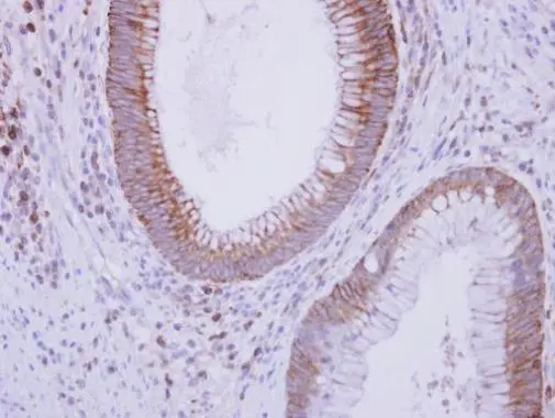
ApplicationsWestern Blot, ELISA
Product group Antibodies
ReactivityHuman, Mouse
TargetC9
Overview
- SupplierBoster Bio
- Product NameAnti-Complement C9 Antibody Picoband(r)
- Delivery Days Customer9
- ApplicationsWestern Blot, ELISA
- CertificationResearch Use Only
- ClonalityPolyclonal
- Concentration500 ug/ml
- Gene ID735
- Target nameC9
- Target descriptioncomplement C9
- Target synonymsARMD15, C9D, complement component C9, complement component 9
- HostRabbit
- IsotypeIgG
- Protein IDP02748
- Protein NameComplement component C9
- Scientific DescriptionBoster Bio Anti-Complement C9 Antibody Picoband® catalog # A01010-2. Tested in ELISA, WB applications. This antibody reacts with Human, Mouse. The brand Picoband indicates this is a premium antibody that guarantees superior quality, high affinity, and strong signals with minimal background in Western blot applications. Only our best-performing antibodies are designated as Picoband, ensuring unmatched performance.
- ReactivityHuman, Mouse
- Storage Instruction-20°C,2°C to 8°C
- UNSPSC12352203
Related products
Product group Antibodies
Anti-C9 AntibodyA101659
ApplicationsWestern Blot, ELISA
ReactivityHuman
- SizePrice
Product group Antibodies
Anti-C9 Antibody144-05622
ApplicationsWestern Blot
ReactivityHuman, Mouse
TargetC9
- SizePrice
Product group Antibodies
References
C5b-9 Polyclonal AntibodyBS-2673R
ApplicationsImmunoFluorescence, ImmunoCytoChemistry, ImmunoHistoChemistry, ImmunoHistoChemistry Frozen, ImmunoHistoChemistry Paraffin
ReactivityHuman, Mouse, Rat
TargetC9
- SizePrice
Product group Antibodies
C9 AntibodyCSB-PA001095
ApplicationsWestern Blot, ELISA
ReactivityHuman
TargetC9
- SizePrice
Product group Antibodies

Goat anti-C9 (aa205-216)EB12362
ApplicationsWestern Blot, ELISA
ReactivityHuman
TargetC9
- SizePrice
Product group Antibodies
ApplicationsImmunoPrecipitation, Western Blot, ImmunoCytoChemistry, ImmunoHistoChemistry
TargetC9
- SizePrice
Product group Antibodies

Complement C9 AntibodyLS-C408853
ApplicationsWestern Blot, ImmunoHistoChemistry
ReactivityHuman, Mouse
TargetC9
- SizePrice
Product group Antibodies
Anti-C9 AntibodyHPA029577
ApplicationsImmunoCytoChemistry, ImmunoHistoChemistry
ReactivityHuman
TargetC9
- SizePrice
Product group Antibodies
C9 antibody [N2C2], InternalGTX109952
ApplicationsWestern Blot, ImmunoHistoChemistry, ImmunoHistoChemistry Paraffin
ReactivityHuman
TargetC9
- SizePrice
Product group Antibodies
Anti-C9 AntibodyCAB5622
ApplicationsWestern Blot, ELISA
ReactivityHuman
TargetC9
- SizePrice