

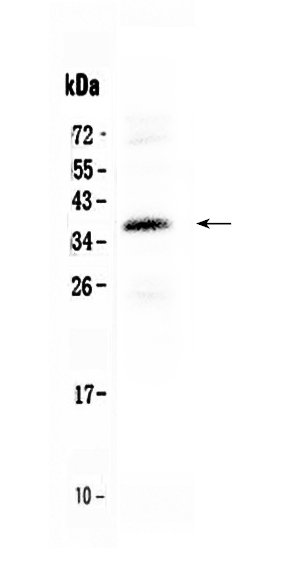
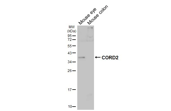
Figure 1. Western blot analysis of CORD2 using anti-CORD2 antibody (A02202-1). Electrophoresis was performed on a 5-20% SDS-PAGE gel at 70V (Stacking gel) / 90V (Resolving gel) for 2-3 hours. The sample well of each lane was loaded with 50ug of sample under reducing conditions. Lane 1: HEPG2 whole cell lysates. After Electrophoresis, proteins were transferred to a Nitrocellulose membrane at 150mA for 50-90 minutes. Blocked the membrane with 5% Non-fat Milk/ TBS for 1.5 hour at RT. The membrane was incubated with rabbit anti-CORD2 antigen affinity purified polyclonal antibody (Catalog # A02202-1) at 0.5 microg/mL overnight at 4°C, then washed with TBS-0.1%Tween 3 times with 5 minutes each and probed with a goat anti-rabbit IgG-HRP secondary antibody at a dilution of 1:10000 for 1.5 hour at RT. The signal is developed using an Enhanced Chemiluminescent detection (ECL) kit (Catalog # EK1002) with Tanon 5200 system. A specific band was detected for CORD2 at approximately 37KD. The expected band size for CORD2 is at 32KD.
Figure 1. Western blot analysis of CORD2 using anti-CORD2 antibody (A02202-1). Electrophoresis was performed on a 5-20% SDS-PAGE gel at 70V (Stacking gel) / 90V (Resolving gel) for 2-3 hours. The sample well of each lane was loaded with 50ug of sample under reducing conditions. Lane 1: HEPG2 whole cell lysates. After Electrophoresis, proteins were transferred to a Nitrocellulose membrane at 150mA for 50-90 minutes. Blocked the membrane with 5% Non-fat Milk/ TBS for 1.5 hour at RT. The membrane was incubated with rabbit anti-CORD2 antigen affinity purified polyclonal antibody (Catalog # A02202-1) at 0.5 microg/mL overnight at 4°C, then washed with TBS-0.1%Tween 3 times with 5 minutes each and probed with a goat anti-rabbit IgG-HRP secondary antibody at a dilution of 1:10000 for 1.5 hour at RT. The signal is developed using an Enhanced Chemiluminescent detection (ECL) kit (Catalog # EK1002) with Tanon 5200 system. A specific band was detected for CORD2 at approximately 37KD. The expected band size for CORD2 is at 32KD.
Anti-CORD2/CRX Antibody Picoband(r)
A02202-1-CARRIER-FREE
ApplicationsWestern Blot
Product group Antibodies
ReactivityHuman
TargetCRX
Overview
- SupplierBoster Bio
- Product NameAnti-CORD2/CRX Antibody Picoband(r)
- Delivery Days Customer9
- ApplicationsWestern Blot
- CertificationResearch Use Only
- ClonalityPolyclonal
- Concentration500 ug/ml
- Gene ID1406
- Target nameCRX
- Target descriptioncone-rod homeobox
- Target synonymsCORD2, CRD, LCA7, OTX3, cone-rod homeobox protein, orthodenticle homeobox 3
- HostRabbit
- IsotypeIgG
- Protein IDO43186
- Protein NameCone-rod homeobox protein
- Scientific DescriptionBoster Bio Anti-CORD2/CRX Antibody Picoband® catalog # A02202-1. Tested in WB applications. This antibody reacts with Human. The brand Picoband indicates this is a premium antibody that guarantees superior quality, high affinity, and strong signals with minimal background in Western blot applications. Only our best-performing antibodies are designated as Picoband, ensuring unmatched performance.
- ReactivityHuman
- Storage Instruction-20°C,2°C to 8°C
- UNSPSC12352203
Related products
Product group Antibodies
Anti-CRX Antibody144-65975
ApplicationsWestern Blot
ReactivityHuman, Mouse, Rat
TargetCRX
- SizePrice
Product group Antibodies
Anti-CRX AntibodyA14932
ApplicationsImmunoFluorescence, Western Blot, ImmunoCytoChemistry
ReactivityMouse
- SizePrice
Product group Antibodies
CRX1 Polyclonal AntibodyBS-3798R
ApplicationsImmunoFluorescence, ELISA, ImmunoCytoChemistry, ImmunoHistoChemistry, ImmunoHistoChemistry Frozen, ImmunoHistoChemistry Paraffin
ReactivityBovine, Canine, Equine, Human, Mouse, Porcine, Rabbit, Rat
TargetCRX
- SizePrice
Product group Antibodies
CRX AntibodyCSB-PA006004DSR1HU
ApplicationsELISA, ImmunoHistoChemistry
ReactivityHuman
TargetCRX
- SizePrice
Product group Antibodies

ApplicationsWestern Blot, ELISA, ImmunoHistoChemistry
ReactivityBovine, Human, Mouse, Porcine, Rat
TargetCRX
- SizePrice
Product group Antibodies

CRD / CRX AntibodyLS-C335596
ApplicationsWestern Blot, ImmunoHistoChemistry
ReactivityHuman, Mouse, Rat
TargetCRX
- SizePrice
Product group Antibodies
CORD2 antibodyGTX124188
ApplicationsWestern Blot, ImmunoHistoChemistry, ImmunoHistoChemistry Frozen, ImmunoHistoChemistry Paraffin
ReactivityHuman, Mouse
TargetCRX
- SizePrice

Product group Antibodies
Anti-CRX AntibodyHPA036763
ApplicationsWestern Blot, ImmunoHistoChemistry
ReactivityHuman
TargetCRX
- SizePrice
Product group Antibodies
Anti-CRX AntibodyCAB5719
ApplicationsImmunoFluorescence, Western Blot, ELISA, ImmunoCytoChemistry
ReactivityHuman
TargetCRX
- SizePrice