

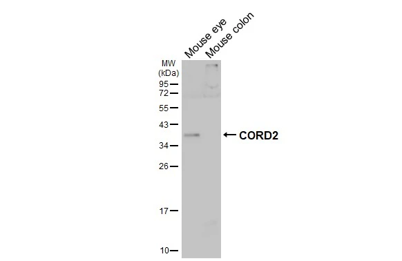
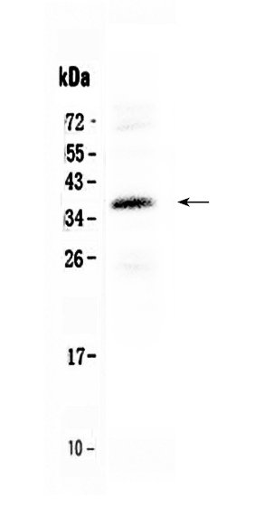
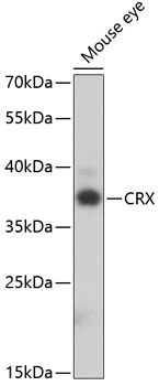
Various tissue extracts (50 μg) were separated by 12% SDS-PAGE, and the membrane was blotted with CORD2 antibody (GTX124188) diluted at 1:1000. The HRP-conjugated anti-rabbit IgG antibody (GTX213110-01) was used to detect the primary antibody.
![CORD2 antibody detects CORD2 protein by immunohistochemical analysis. Sample: Frozen-sectioned mouse eye. Green: CORD2 stained by CORD2 antibody (GTX124188) diluted at 1:500. Red: beta Tubulin 3/ Tuj1 antibody [GT11710] (GTX631836) diluted at 1:500. Blue: Hoechst 33342 staining.](https://www.genetex.com/upload/website/prouct_img/normal/GTX124188/GTX124188_45708_20250620_IHC-Fr_M_25062520_221.webp "CORD2 antibody detects CORD2 protein by immunohistochemical analysis. Sample: Frozen-sectioned mouse eye. Green: CORD2 stained by CORD2 antibody (GTX124188) diluted at 1:500. Red: beta Tubulin 3/ Tuj1 antibody [GT11710] (GTX631836) diluted at 1:500. Blue: Hoechst 33342 staining.")
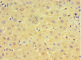
![CORD2 antibody detects CORD2 protein by immunohistochemical analysis. Sample: Paraffin-embedded mouse eye. Green: CORD2 stained by CORD2 antibody (GTX124188) diluted at 1:1250. Red: beta Tubulin 3/ Tuj1 antibody [GT11710] (GTX631836) diluted at 1:500. Blue: Fluoroshield with DAPI (GTX30920). Antigen Retrieval: Citrate buffer, pH 6.0, 15 min](https://www.genetex.com/upload/website/prouct_img/normal/GTX124188/GTX124188_45708_20250620_IHC-P_M_25062520_411.webp "CORD2 antibody detects CORD2 protein by immunohistochemical analysis. Sample: Paraffin-embedded mouse eye. Green: CORD2 stained by CORD2 antibody (GTX124188) diluted at 1:1250. Red: beta Tubulin 3/ Tuj1 antibody [GT11710] (GTX631836) diluted at 1:500. Blue: Fluoroshield with DAPI (GTX30920). Antigen Retrieval: Citrate buffer, pH 6.0, 15 min")
Various tissue extracts (50 μg) were separated by 12% SDS-PAGE, and the membrane was blotted with CORD2 antibody (GTX124188) diluted at 1:1000. The HRP-conjugated anti-rabbit IgG antibody (GTX213110-01) was used to detect the primary antibody.
CORD2 antibody
GTX124188
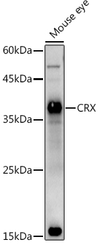
ApplicationsWestern Blot, ImmunoHistoChemistry, ImmunoHistoChemistry Frozen, ImmunoHistoChemistry Paraffin
Product group Antibodies
ReactivityHuman, Mouse
TargetCRX
Overview
- SupplierGeneTex
- Product NameCORD2 antibody
- Delivery Days Customer9
- Application Supplier NoteWB: 1:500-1:3000. IHC-P: 1:100-1:1000. *Optimal dilutions/concentrations should be determined by the researcher.Not tested in other applications.
- ApplicationsWestern Blot, ImmunoHistoChemistry, ImmunoHistoChemistry Frozen, ImmunoHistoChemistry Paraffin
- CertificationResearch Use Only
- ClonalityPolyclonal
- Concentration0.85 mg/ml
- ConjugateUnconjugated
- Gene ID1406
- Target nameCRX
- Target descriptioncone-rod homeobox
- Target synonymsCORD2, CRD, LCA7, OTX3, cone-rod homeobox protein, orthodenticle homeobox 3
- HostRabbit
- IsotypeIgG
- Protein IDO43186
- Protein NameCone-rod homeobox protein
- Scientific DescriptionThe protein encoded by this gene is a photoreceptor-specific transcription factor which plays a role in the differentiation of photoreceptor cells. This homeodomain protein is necessary for the maintenance of normal cone and rod function. Mutations in this gene are associated with photoreceptor degeneration, Leber congenital amaurosis type III and the autosomal dominant cone-rod dystrophy 2. Several alternatively spliced transcript variants of this gene have been described, but the full-length nature of some variants has not been determined. [provided by RefSeq]
- ReactivityHuman, Mouse
- Storage Instruction-20°C or -80°C,2°C to 8°C
- UNSPSC41116161
Datasheet
Related products
Product group Antibodies
Anti-CRX Antibody144-65975
ApplicationsWestern Blot
ReactivityHuman, Mouse, Rat
TargetCRX
- SizePrice
Product group Antibodies
Anti-CRX AntibodyA14932
ApplicationsImmunoFluorescence, Western Blot, ImmunoCytoChemistry
ReactivityMouse
- SizePrice
Product group Antibodies
Anti-CORD2/CRX Antibody Picoband(r)A02202-1-CARRIER-FREE
ApplicationsWestern Blot
ReactivityHuman
TargetCRX
- SizePrice
Product group Antibodies
CRX1 Polyclonal AntibodyBS-3798R
ApplicationsImmunoFluorescence, ELISA, ImmunoCytoChemistry, ImmunoHistoChemistry, ImmunoHistoChemistry Frozen, ImmunoHistoChemistry Paraffin
ReactivityBovine, Canine, Equine, Human, Mouse, Porcine, Rabbit, Rat
TargetCRX
- SizePrice
Product group Antibodies
CRX AntibodyCSB-PA006004DSR1HU
ApplicationsELISA, ImmunoHistoChemistry
ReactivityHuman
TargetCRX
- SizePrice
Product group Antibodies

ApplicationsWestern Blot, ELISA, ImmunoHistoChemistry
ReactivityBovine, Human, Mouse, Porcine, Rat
TargetCRX
- SizePrice
Product group Antibodies

CRD / CRX AntibodyLS-C335596
ApplicationsWestern Blot, ImmunoHistoChemistry
ReactivityHuman, Mouse, Rat
TargetCRX
- SizePrice
Product group Antibodies
CORD2 antibodyGTX33111
ApplicationsWestern Blot
ReactivityHuman, Mouse
TargetCRX
- SizePrice

Product group Antibodies
Anti-CRX AntibodyHPA036763
ApplicationsWestern Blot, ImmunoHistoChemistry
ReactivityHuman
TargetCRX
- SizePrice