


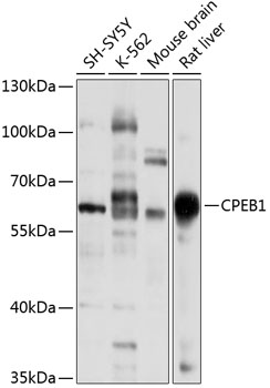
Figure 1. Western blot analysis of CPEB1 using anti-CPEB1 antibody (M03578). Electrophoresis was performed on a 5-20% SDS-PAGE gel at 70V (Stacking gel) / 90V (Resolving gel) for 2-3 hours. The sample well of each lane was loaded with 30 ug of sample under reducing conditions. Lane 1: human A549 whole cell lysates, Lane 2: human K562 whole cell lysates, Lane 3: human SiHa whole cell lysates, Lane 4: human U251 whole cell lysates. After electrophoresis, proteins were transferred to a nitrocellulose membrane at 150 mA for 50-90 minutes. Blocked the membrane with 5% non-fat milk/TBS for 1.5 hour at RT. The membrane was incubated with rabbit anti-CPEB1 antigen affinity purified monoclonal antibody (Catalog # M03578) at 1:1000 overnight at 4°C, then washed with TBS-0.1%Tween 3 times with 5 minutes each and probed with a goat anti-rabbit IgG-HRP secondary antibody at a dilution of 1:500 for 1.5 hour at RT. The signal is developed using an Enhanced Chemiluminescent detection (ECL) kit (Catalog # EK1002) with Tanon 5200 system. A specific band was detected for CPEB1 at approximately 70 kDa. The expected band size for CPEB1 is at 63 kDa.
Figure 1. Western blot analysis of CPEB1 using anti-CPEB1 antibody (M03578). Electrophoresis was performed on a 5-20% SDS-PAGE gel at 70V (Stacking gel) / 90V (Resolving gel) for 2-3 hours. The sample well of each lane was loaded with 30 ug of sample under reducing conditions. Lane 1: human A549 whole cell lysates, Lane 2: human K562 whole cell lysates, Lane 3: human SiHa whole cell lysates, Lane 4: human U251 whole cell lysates. After electrophoresis, proteins were transferred to a nitrocellulose membrane at 150 mA for 50-90 minutes. Blocked the membrane with 5% non-fat milk/TBS for 1.5 hour at RT. The membrane was incubated with rabbit anti-CPEB1 antigen affinity purified monoclonal antibody (Catalog # M03578) at 1:1000 overnight at 4°C, then washed with TBS-0.1%Tween 3 times with 5 minutes each and probed with a goat anti-rabbit IgG-HRP secondary antibody at a dilution of 1:500 for 1.5 hour at RT. The signal is developed using an Enhanced Chemiluminescent detection (ECL) kit (Catalog # EK1002) with Tanon 5200 system. A specific band was detected for CPEB1 at approximately 70 kDa. The expected band size for CPEB1 is at 63 kDa.
Anti-CPEB1 Rabbit Monoclonal Antibody
M03578
ApplicationsImmunoPrecipitation, Western Blot
Product group Antibodies
ReactivityHuman
TargetCPEB1
Overview
- SupplierBoster Bio
- Product NameAnti-CPEB1 Rabbit Monoclonal Antibody
- Delivery Days Customer9
- ApplicationsImmunoPrecipitation, Western Blot
- CertificationResearch Use Only
- ClonalityMonoclonal
- Clone ID22C36
- Gene ID64506
- Target nameCPEB1
- Target descriptioncytoplasmic polyadenylation element binding protein 1
- Target synonymsCPE-BP1, CPEB, CPEB-1, h-CPEB, hCPEB-1, cytoplasmic polyadenylation element-binding protein 1, CPE-binding protein 1
- HostRabbit
- IsotypeIgG
- Protein IDQ9BZB8
- Protein NameCytoplasmic polyadenylation element-binding protein 1
- Scientific DescriptionBoster Bio Anti-CPEB1 Rabbit Monoclonal Antibody catalog # M03578. Tested in WB, IP applications. This antibody reacts with Human.
- ReactivityHuman
- Storage Instruction-20°C
- UNSPSC12352203
Related products
Product group Antibodies
Anti-CPEB1 AntibodyA15048
ApplicationsWestern Blot
ReactivityHuman, Mouse, Rat
- SizePrice
Product group Antibodies
CPEB1 Polyclonal AntibodyBS-6037R
ApplicationsImmunoFluorescence, Western Blot, ELISA, ImmunoCytoChemistry, ImmunoHistoChemistry, ImmunoHistoChemistry Frozen, ImmunoHistoChemistry Paraffin
ReactivityBovine, Chicken, Equine, Human, Mouse, Porcine, Rabbit, Rat
TargetCPEB1
- SizePrice
Product group Antibodies
CPEB1 AntibodyCSB-PA861184LA01HU
ApplicationsELISA
ReactivityHuman
TargetCPEB1
- SizePrice
Product group Antibodies

Goat anti-CPEB1EB06391
ApplicationsWestern Blot, ELISA
ReactivityHuman
TargetCPEB1
- SizePrice
Product group Antibodies
ApplicationsImmunoPrecipitation, Western Blot, ImmunoCytoChemistry, ImmunoHistoChemistry
ReactivityMouse, Rat
TargetCPEB1
- SizePrice
Product group Antibodies

CPEB1 / CPEB Antibody (aa1-486)LS-C371992
ApplicationsELISA
ReactivityHuman
TargetCPEB1
- SizePrice
Product group Antibodies
Anti-CPEB1 AntibodyHPA040396
ApplicationsImmunoCytoChemistry
ReactivityHuman
TargetCPEB1
- SizePrice
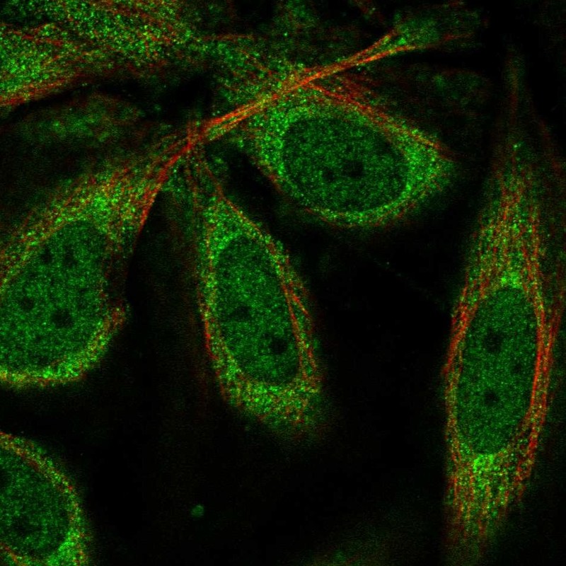
![CPEB1 antibody [C3], C-term detects CPEB1 protein by immunofluorescent analysis. Sample: DIV9 rat E18 primary hippocampal neuron cells were fixed in 4% paraformaldehyde at RT for 15 min. Green: CPEB1 stained by CPEB1 antibody [C3], C-term (GTX104682) diluted at 1:500. Red: beta Tubulin 3/ Tuj1, stained by beta Tubulin 3/ Tuj1 antibody [GT11710] (GTX631836) diluted at 1:500. Blue: Fluoroshield with DAPI (GTX30920).](https://www.genetex.com/upload/website/prouct_img/normal/GTX104682/GTX104682_39820_20181004_ICC_IF_R_w_23060120_107.webp)
Product group Antibodies
CPEB1 antibody [C3], C-termGTX104682
ApplicationsImmunoFluorescence, Western Blot, ImmunoCytoChemistry
ReactivityHuman
TargetCPEB1
- SizePrice