


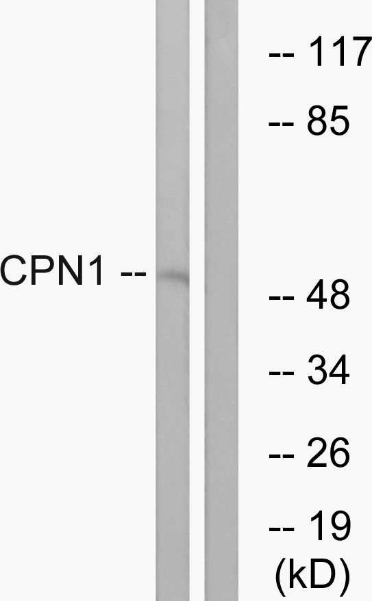
Figure 1. Western blot analysis of CPN1 using anti-CPN1 antibody (A05704-2). Electrophoresis was performed on a 5-20% SDS-PAGE gel at 70V (Stacking gel) / 90V (Resolving gel) for 2-3 hours. The sample well of each lane was loaded with 30ug of sample under reducing conditions. Lane 1: rat liver tissue lysates, Lane 2: mouse liver tissue lysates, Lane 3: rat C6 whole cell lysates, Lane 4: mouse HEPA1-6 whole cell lysates. After Electrophoresis, proteins were transferred to a Nitrocellulose membrane at 150mA for 50-90 minutes. Blocked the membrane with 5% Non-fat Milk/ TBS for 1.5 hour at RT. The membrane was incubated with rabbit anti-CPN1 antigen affinity purified polyclonal antibody (Catalog # A05704-2) at 0.5 microg/mL overnight at 4°C, then washed with TBS-0.1%Tween 3 times with 5 minutes each and probed with a goat anti-rabbit IgG-HRP secondary antibody at a dilution of 1:5000 for 1.5 hour at RT. The signal is developed using an Enhanced Chemiluminescent detection (ECL) kit (Catalog # EK1002) with Tanon 5200 system. A specific band was detected for CPN1 at approximately 52KD. The expected band size for CPN1 is at 52KD.
. CPN1 was detected in immunocytochemical section of CACO-2 cells. Enzyme antigen retrieval was performed using IHC enzyme antigen retrieval reagent (AR0022) for 15 mins. The cells were blocked with 10% goat serum. And then incubated with 5microg/mL rabbit anti-CPN1 Antibody (A05704-2) overnight at 4°C. DyLight®488 Conjugated Goat Anti-Rabbit IgG (BA1127) was used as secondary antibody at 1:100 dilution and incubated for 30 minutes at 37°C. The section was counterstained with DAPI. Visualize using a fluorescence microscope and filter sets appropriate for the label used.")
. Overlay histogram showing HepG2 cells stained with A05704-2 (Blue line). The cells were fixed with 4% paraformaldehyde and blocked with 10% normal goat serum. And then incubated with rabbit anti-CPN1 Antibody (A05704-2, 1microg/1x106 cells) for 30 min at 20°C. DyLight®488 conjugated goat anti-rabbit IgG (BA1127, 5-10microg/1x106 cells) was used as secondary antibody for 30 minutes at 20°C. Isotype control antibody (Green line) was rabbit IgG (1microg/1x106) used under the same conditions. Unlabelled sample without incubation with primary antibody and secondary antibody (Red line) was used as a blank control.")
Figure 1. Western blot analysis of CPN1 using anti-CPN1 antibody (A05704-2). Electrophoresis was performed on a 5-20% SDS-PAGE gel at 70V (Stacking gel) / 90V (Resolving gel) for 2-3 hours. The sample well of each lane was loaded with 30ug of sample under reducing conditions. Lane 1: rat liver tissue lysates, Lane 2: mouse liver tissue lysates, Lane 3: rat C6 whole cell lysates, Lane 4: mouse HEPA1-6 whole cell lysates. After Electrophoresis, proteins were transferred to a Nitrocellulose membrane at 150mA for 50-90 minutes. Blocked the membrane with 5% Non-fat Milk/ TBS for 1.5 hour at RT. The membrane was incubated with rabbit anti-CPN1 antigen affinity purified polyclonal antibody (Catalog # A05704-2) at 0.5 microg/mL overnight at 4°C, then washed with TBS-0.1%Tween 3 times with 5 minutes each and probed with a goat anti-rabbit IgG-HRP secondary antibody at a dilution of 1:5000 for 1.5 hour at RT. The signal is developed using an Enhanced Chemiluminescent detection (ECL) kit (Catalog # EK1002) with Tanon 5200 system. A specific band was detected for CPN1 at approximately 52KD. The expected band size for CPN1 is at 52KD.
Anti-CPN1 Antibody Picoband(r)
A05704-2-DYLIGHT594
ApplicationsFlow Cytometry, ImmunoFluorescence, Western Blot, ELISA, ImmunoCytoChemistry
Product group Antibodies
ReactivityHuman, Mouse, Rat
TargetCPN1
Overview
- SupplierBoster Bio
- Product NameAnti-CPN1 Antibody Picoband(r)
- Delivery Days Customer9
- ApplicationsFlow Cytometry, ImmunoFluorescence, Western Blot, ELISA, ImmunoCytoChemistry
- CertificationResearch Use Only
- ClonalityPolyclonal
- Concentration500 ug/ml
- ConjugateOther Conjugate
- Gene ID1369
- Target nameCPN1
- Target descriptioncarboxypeptidase N subunit 1
- Target synonymsCPN, SCPN, carboxypeptidase N catalytic chain, anaphylatoxin inactivator, arginine carboxypeptidase, carboxypeptidase K, carboxypeptidase N catalytic subunit, carboxypeptidase N polypeptide 1 50 kD, carboxypeptidase N small subunit, carboxypeptidase N, polypeptide 1, kininase I, kininase-1, lysine carboxypeptidase, plasma carboxypeptidase B, serum carboxypeptidase N
- HostRabbit
- IsotypeIgG
- Protein IDP15169
- Protein NameCarboxypeptidase N catalytic chain
- Scientific DescriptionBoster Bio Anti-CPN1 Antibody Picoband® catalog # A05704-2. Tested in ELISA, Flow Cytometry, IF, ICC, WB applications. This antibody reacts with Human, Mouse, Rat. The brand Picoband indicates this is a premium antibody that guarantees superior quality, high affinity, and strong signals with minimal background in Western blot applications. Only our best-performing antibodies are designated as Picoband, ensuring unmatched performance.
- ReactivityHuman, Mouse, Rat
- Storage Instruction-20°C,2°C to 8°C
- UNSPSC12352203
Related products
Product group Antibodies
Cpn1 Polyclonal AntibodyCAC10789
ApplicationsWestern Blot, ELISA, ImmunoHistoChemistry
ReactivityMouse
TargetCPN1
- SizePrice
Product group Antibodies
Anti-CPN1 (Center) Antibody102-21265
ApplicationsFlow Cytometry, Western Blot, ImmunoHistoChemistry, ImmunoHistoChemistry Paraffin
TargetCPN1
- SizePrice
Product group Antibodies
Anti-CPN1 AntibodyA101485
ApplicationsWestern Blot, ELISA
ReactivityHuman
- SizePrice
Product group Antibodies
CPN1 antibody [N1C3]GTX105515
ApplicationsWestern Blot
ReactivityHuman
TargetCPN1
- SizePrice
Product group Antibodies

CPN1 AntibodyLS-C409436
ApplicationsWestern Blot
ReactivityHuman, Mouse, Rat
TargetCPN1
- SizePrice
Product group Antibodies
CPN1 AntibodyCSB-PA001774
ApplicationsWestern Blot, ELISA
ReactivityHuman
TargetCPN1
- SizePrice
Product group Antibodies
Anti-CPN1 Antibody Picoband(r)A05704-2-CARRIER-FREE
ApplicationsFlow Cytometry, ImmunoFluorescence, Western Blot, ELISA, ImmunoCytoChemistry
ReactivityHuman, Mouse, Rat
TargetCPN1
- SizePrice