


Immunohistochemical staining of human retina shows cytoplasmic positivity in cells in inner nuclear layer and nerve fibers.
Immunohistochemical staining of human retina shows cytoplasmic positivity in cells in inner nuclear layer and nerve fibers.
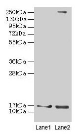
Anti-CRABP1 Antibody
HPA017203
ApplicationsImmunoHistoChemistry
Product group Antibodies
ReactivityHuman
TargetCRABP1
Overview
- SupplierAtlas Antibodies
- Product NameAnti-CRABP1 Antibody
- Delivery Days Customer4
- ApplicationsImmunoHistoChemistry
- CertificationResearch Use Only
- ClonalityPolyclonal
- ConjugateUnconjugated
- Gene ID1381
- Target nameCRABP1
- Target descriptioncellular retinoic acid binding protein 1
- Target synonymsCRABP, CRABP-I, CRABPI, RBP5, cellular retinoic acid-binding protein 1, cellular retinoic acid-binding protein I
- HostRabbit
- IsotypeIgG
- Protein IDP29762
- Protein NameCellular retinoic acid-binding protein 1
- Scientific DescriptionRecombinant Protein Epitope Signature Tag (PrEST) antigen sequence
- ReactivityHuman
- Storage Instruction-20°C,2°C to 8°C
- UNSPSC41116161
Datasheet
MSDS
Related products
Product group Antibodies
Anti-CRABP1 (C-term) Antibody102-26836
ApplicationsWestern Blot, ImmunoHistoChemistry, ImmunoHistoChemistry Paraffin
TargetCRABP1
- SizePrice

Product group Antibodies
Anti-CRABP1 Antibody Picoband(r)A07951-1-CARRIER-FREE
ApplicationsFlow Cytometry, Western Blot, ELISA
ReactivityHuman
TargetCRABP1
- SizePrice
Product group Antibodies

CRABP1 / CRABP AntibodyLS-C830465
ApplicationsWestern Blot, ELISA, ImmunoHistoChemistry
ReactivityHuman, Mouse, Rat
TargetCRABP1
- SizePrice
Product group Antibodies
CRABP1 Recombinant AntibodyBSM-62149R
ApplicationsImmunoPrecipitation, Western Blot
ReactivityHuman, Mouse
TargetCRABP1
- SizePrice
Product group Antibodies
Crabp1 Polyclonal AntibodyCAC08331
ApplicationsWestern Blot, ELISA, ImmunoHistoChemistry
ReactivityMouse
TargetCRABP1
- SizePrice
Product group Antibodies
CRABP1 AntibodyCSB-PA005935DA01HU
ApplicationsWestern Blot, ELISA, ImmunoHistoChemistry
ReactivityHuman, Mouse
TargetCRABP1
- SizePrice
![ICC/IF analysis of 3T3 cells using GTX50013 CRABP1 antibody [AT1A1]. Dilution : 1:100](https://www.genetex.com/upload/website/prouct_img/normal/GTX50013/GTX50013_20231002_ICCIF_23100123_587.webp)
Product group Antibodies
CRABP1 antibody [AT1A1]GTX50013
ApplicationsFlow Cytometry, ImmunoFluorescence, Western Blot, ELISA, ImmunoCytoChemistry
ReactivityHuman, Mouse
TargetCRABP1
- SizePrice