


Immunofluorescent staining of human cell line PC-3 shows localization to nucleus & nucleoli.
Immunofluorescent staining of human cell line PC-3 shows localization to nucleus & nucleoli.
Anti-CUL1 Antibody
HPA064584
ApplicationsImmunoCytoChemistry
Product group Antibodies
ReactivityHuman
TargetCUL1
Overview
- SupplierAtlas Antibodies
- Product NameAnti-CUL1 Antibody
- Delivery Days Customer4
- ApplicationsImmunoCytoChemistry
- CertificationResearch Use Only
- ClonalityPolyclonal
- ConjugateUnconjugated
- Gene ID8454
- Target nameCUL1
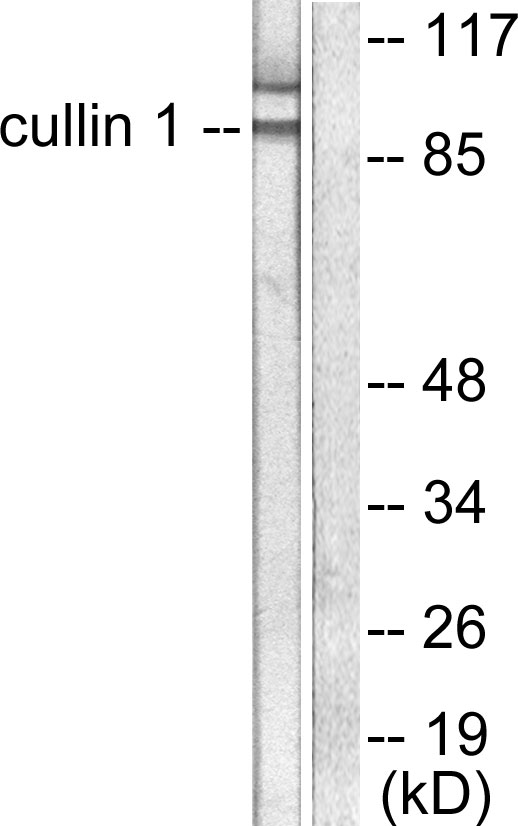
- Target descriptioncullin 1
- Target synonymscullin-1, CUL-1
- HostRabbit
- IsotypeIgG
- Protein IDQ13616
- Protein NameCullin-1
- Scientific DescriptionRecombinant Protein Epitope Signature Tag (PrEST) antigen sequence
- ReactivityHuman
- Storage Instruction-20°C,2°C to 8°C
- UNSPSC41116161
Datasheet
MSDS
Related products
Product group Antibodies
Anti-Cullin 1 AntibodyA95442
ApplicationsImmunoFluorescence, Western Blot, ELISA, ImmunoHistoChemistry
ReactivityHuman, Mouse
- SizePrice
Product group Antibodies
Anti-CUL1 (C-term) Antibody102-22261
ApplicationsWestern Blot
TargetCUL1
- SizePrice
Product group Antibodies
Cullin 1 Recombinant Antibody, AbBy Fluor-594 ConjugatedBSM-61690R-BF594
ApplicationsFlow Cytometry, ImmunoFluorescence, Western Blot
ReactivityHuman, Mouse, Rat
TargetCUL1
- SizePrice
Product group Antibodies
CUL1 AntibodyCSB-PA001830
ApplicationsImmunoFluorescence, Western Blot, ELISA, ImmunoHistoChemistry
ReactivityHuman, Mouse
TargetCUL1
- SizePrice
Product group Antibodies
ApplicationsImmunoPrecipitation, Western Blot, ImmunoCytoChemistry, ImmunoHistoChemistry
ReactivityMouse, Rat
TargetCUL1
- SizePrice
![Various whole cell extracts (30 μg) were separated by 7.5% SDS-PAGE, and the membrane was blotted with Cullin 1 antibody [JM72-30] (GTX00988) diluted at 1:500. The HRP-conjugated anti-rabbit IgG antibody (GTX213110-01) was used to detect the primary antibody.](https://www.genetex.com/upload/website/prouct_img/normal/GTX00988/GTX00988_HK0904_20200214_WB_w_23053121_877.webp)
Product group Antibodies
Cullin 1 antibody [JM72-30]GTX00988
ApplicationsFlow Cytometry, Western Blot, ImmunoHistoChemistry, ImmunoHistoChemistry Paraffin
ReactivityHuman, Mouse, Rat
TargetCUL1
- SizePrice
Product group Antibodies

CUL1 / Cullin 1 AntibodyLS-C331929
ApplicationsImmunoFluorescence, Western Blot, ImmunoHistoChemistry
ReactivityHuman, Mouse, Rat
TargetCUL1
- SizePrice
Product group Antibodies
Anti-CUL1Y058318
ApplicationsWestern Blot, ELISA, ImmunoHistoChemistry
ReactivityHuman, Mouse, Rat
- SizePrice