


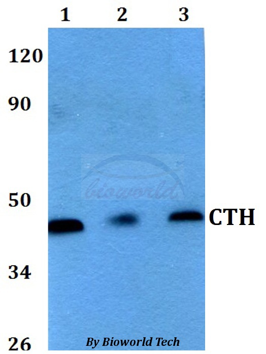
Figure 1. Western blot analysis of CTH using anti-CTH antibody (PA1556). Electrophoresis was performed on a 5-20% SDS-PAGE gel at 70V (Stacking gel) / 90V (Resolving gel) for 2-3 hours. The sample well of each lane was loaded with 30 ug of sample under reducing conditions. Lane 1: human K562 whole cell lysates, Lane 2: human HepG2 whole cell lysates, Lane 3: human THP-1 whole cell lysates, Lane 4: human HCCT tissue lysates, Lane 5: rat liver tissue lysates, Lane 6: mouse liver tissue lysates. After electrophoresis, proteins were transferred to a nitrocellulose membrane at 150 mA for 50-90 minutes. Blocked the membrane with 5% non-fat milk/TBS for 1.5 hour at RT. The membrane was incubated with rabbit anti-CTH antigen affinity purified polyclonal antibody (Catalog # PA1556) at 0.5 microg/mL overnight at 4°C, then washed with TBS-0.1%Tween 3 times with 5 minutes each and probed with a goat anti-rabbit IgG-HRP secondary antibody at a dilution of 1:5000 for 1.5 hour at RT. The signal is developed using an Enhanced Chemiluminescent detection (ECL) kit (Catalog # EK1002) with Tanon 5200 system. A specific band was detected for CTH at approximately 45 kDa. The expected band size for CTH is at 45 kDa.
. CTH was detected in an immunocytochemical section of A431 cells. Enzyme antigen retrieval was performed using IHC enzyme antigen retrieval reagent (AR0022) for 15 mins. The cells were blocked with 10% goat serum. And then incubated with 5 microg/mL rabbit anti-CTH Antibody (PA1556) overnight at 4°C. DyLight®488 Conjugated Goat Anti-Rabbit IgG (BA1127) was used as secondary antibody at 1:100 dilution and incubated for 30 minutes at 37°C. The section was counterstained with DAPI. Visualize using a fluorescence microscope and filter sets appropriate for the label used.")
. Overlay histogram showing JK cells stained with PA1556 (Blue line). To facilitate intracellular staining, cells were fixed with 4% paraformaldehyde and permeabilized with permeabilization buffer. The cells were blocked with 10% normal goat serum. And then incubated with rabbit anti-CTH Antibody (PA1556, 1 microg/1x106 cells) for 30 min at 20°C. DyLight®488 conjugated goat anti-rabbit IgG (BA1127, 5-10 microg/1x106 cells) was used as secondary antibody for 30 minutes at 20°C. Isotype control antibody (Green line) was rabbit IgG (1 microg/1x106) used under the same conditions. Unlabelled sample without incubation with primary antibody and secondary antibody (Red line) was used as a blank control.")
Figure 1. Western blot analysis of CTH using anti-CTH antibody (PA1556). Electrophoresis was performed on a 5-20% SDS-PAGE gel at 70V (Stacking gel) / 90V (Resolving gel) for 2-3 hours. The sample well of each lane was loaded with 30 ug of sample under reducing conditions. Lane 1: human K562 whole cell lysates, Lane 2: human HepG2 whole cell lysates, Lane 3: human THP-1 whole cell lysates, Lane 4: human HCCT tissue lysates, Lane 5: rat liver tissue lysates, Lane 6: mouse liver tissue lysates. After electrophoresis, proteins were transferred to a nitrocellulose membrane at 150 mA for 50-90 minutes. Blocked the membrane with 5% non-fat milk/TBS for 1.5 hour at RT. The membrane was incubated with rabbit anti-CTH antigen affinity purified polyclonal antibody (Catalog # PA1556) at 0.5 microg/mL overnight at 4°C, then washed with TBS-0.1%Tween 3 times with 5 minutes each and probed with a goat anti-rabbit IgG-HRP secondary antibody at a dilution of 1:5000 for 1.5 hour at RT. The signal is developed using an Enhanced Chemiluminescent detection (ECL) kit (Catalog # EK1002) with Tanon 5200 system. A specific band was detected for CTH at approximately 45 kDa. The expected band size for CTH is at 45 kDa.
Anti-Cystathionase/CTH Antibody Picoband(r)
PA1556
ApplicationsFlow Cytometry, ImmunoFluorescence, Western Blot, ImmunoCytoChemistry
Product group Antibodies
ReactivityHuman, Mouse, Rabbit, Rat
TargetCTH
Overview
- SupplierBoster Bio
- Product NameAnti-Cystathionase/CTH Antibody Picoband(r)
- Delivery Days Customer9
- Application Supplier NoteTested Species: In-house tested species with positive results. Predicted Species: Species predicted to be fit for the product based on sequence similarities. By Heat: Boiling the paraffin sections in 10mM citrate buffer, pH6.0, for 20mins is required for the staining of formalin/paraffin sections. Other applications have not been tested. Optimal dilutions should be determined by end users.
- ApplicationsFlow Cytometry, ImmunoFluorescence, Western Blot, ImmunoCytoChemistry
- Applications SupplierIHP, WB, IHC
- CertificationResearch Use Only
- ClonalityPolyclonal
- Concentration500 ug/ml
- Gene ID1491
- Target nameCTH
- Target descriptioncystathionine gamma-lyase
- Target synonymsCGL, CSE, cystathionine gamma-lyase, cystathionase (cystathionine gamma-lyase), cysteine desulfhydrase, cysteine-protein sulfhydrase, gamma-cystathionase, homocysteine desulfhydrase, homoserine deaminase, homoserine dehydratase
- HostRabbit
- IsotypeIgG
- Protein IDP32929
- Protein NameCystathionine gamma-lyase
- Scientific DescriptionBoster Bio Anti-Cystathionase/CTH Antibody catalog # PA1556. Tested in Flow Cytometry, IF, ICC, WB applications. This antibody reacts with Human, Mouse, Rat. The brand Picoband indicates this is a premium antibody that guarantees superior quality, high affinity, and strong signals with minimal background in Western blot applications. Only our best-performing antibodies are designated as Picoband, ensuring unmatched performance.
- ReactivityHuman, Mouse, Rabbit, Rat
- Storage Instruction-20°C,2°C to 8°C
- UNSPSC12352203
References
- Matyasova K, Soltysova A, Babula P, et al. Role of the 3-mercaptopyruvate sulfurtransferase in colon/colorectal cancers. Eur J Cell Biol. 2024,103(2):151415. doi: 10.1016/j.ejcb.2024.151415Read this paper
- Yang C, Chen J, Yu Z, et al. Mining of RNA Methylation-Related Genes and Elucidation of Their Molecular Biology in Gallbladder Carcinoma. Front Oncol. 2021,11:621806. doi: 10.3389/fonc.2021.621806Read this paper
- Fan ZG, Qu XL, Chu P, et al. MicroRNA-210 promotes angiogenesis in acute myocardial infarction. Mol Med Rep. 2018,17(4):5658-5665. doi: 10.3892/mmr.2018.8620Read this paper
- Yang Y, Hou J, Shao M, et al. CXCL5 as an autocrine or paracrine cytokine is associated with proliferation and migration of hepatoblastoma HepG2 cells. Oncol Lett. 2017,14(6):7977-7985. doi: 10.3892/ol.2017.7236Read this paper
Datasheet
MSDS
Related products
Product group Antibodies
Anti-CTH AntibodyA28805
ApplicationsWestern Blot
ReactivityHuman, Mouse, Rat
- SizePrice
Product group Antibodies
Anti-CTH Antibody144-06121
ApplicationsWestern Blot
ReactivityHuman, Mouse
TargetCTH
- SizePrice
Product group Antibodies
CTH Polyclonal AntibodyBS-9515R
ApplicationsImmunoFluorescence, Western Blot, ImmunoHistoChemistry, ImmunoHistoChemistry Paraffin
ReactivityBovine, Canine, Human, Mouse, Rabbit, Rat
TargetCTH
- SizePrice
Product group Antibodies
CTH AntibodyCSB-PA006160LA01HU
ApplicationsImmunoFluorescence, Western Blot, ELISA, ImmunoHistoChemistry
ReactivityHuman, Mouse, Rat
TargetCTH
- SizePrice
Product group Antibodies
CTH Polyclonal AntibodyCAC13018
ApplicationsImmunoFluorescence, Western Blot, ELISA, ImmunoHistoChemistry
ReactivityMouse, Rat
TargetCTH
- SizePrice
Product group Antibodies

CTH / Cystathionase AntibodyLS-C334514
ApplicationsWestern Blot, ImmunoHistoChemistry
ReactivityHuman, Mouse, Rat
TargetCTH
- SizePrice

Product group Antibodies
Anti-CTH AntibodyHPA021591
ApplicationsImmunoHistoChemistry
ReactivityHuman
TargetCTH
- SizePrice
Product group Antibodies
CTH antibodyGTX113409
ApplicationsWestern Blot, ImmunoHistoChemistry, ImmunoHistoChemistry Paraffin
ReactivityHuman, Mouse, Rat
TargetCTH
- SizePrice
Product group Antibodies
ApplicationsWestern Blot, ELISA
ReactivityHuman
TargetCTH
- SizePrice