


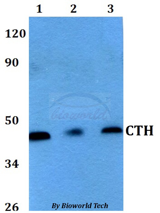
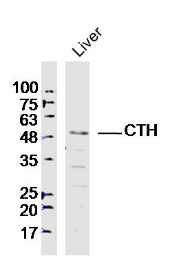
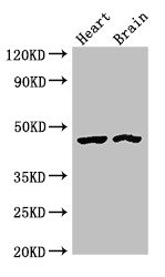
Figure 1. Western blot analysis of Cystathionase using anti-Cystathionase antibody (PB9494). Electrophoresis was performed on a 5-20% SDS-PAGE gel at 70V (Stacking gel) / 90V (Resolving gel) for 2-3 hours. The sample well of each lane was loaded with 50ug of sample under reducing conditions. lane 1: K562 whole cell lysates, lane 2: HepG2 whole cell lysates, lane 3: RH35 whole cell lysates, lane 4: rat liver tissue lysates, lane 5: rat kidney tissue lysates, lane 6: mouse liver tissue lysates. After Electrophoresis, proteins were transferred to a Nitrocellulose membrane at 150mA for 50-90 minutes. Blocked the membrane with 5% Non-fat Milk/ TBS for 1.5 hour at RT. The membrane was incubated with rabbit anti-Cystathionase antigen affinity purified polyclonal antibody (Catalog # PB9494) at 0.5 microg/mL overnight at 4°C, then washed with TBS-0.1%Tween 3 times with 5 minutes each and probed with a goat anti-rabbit IgG-HRP secondary antibody at a dilution of 1:10000 for 1.5 hour at RT. The signal is developed using an Enhanced Chemiluminescent detection (ECL) kit (Catalog # EK1002) with Tanon 5200 system. A specific band was detected for Cystathionase at approximately 45KD. The expected band size for Cystathionase is at 45KD.
Figure 1. Western blot analysis of Cystathionase using anti-Cystathionase antibody (PB9494). Electrophoresis was performed on a 5-20% SDS-PAGE gel at 70V (Stacking gel) / 90V (Resolving gel) for 2-3 hours. The sample well of each lane was loaded with 50ug of sample under reducing conditions. lane 1: K562 whole cell lysates, lane 2: HepG2 whole cell lysates, lane 3: RH35 whole cell lysates, lane 4: rat liver tissue lysates, lane 5: rat kidney tissue lysates, lane 6: mouse liver tissue lysates. After Electrophoresis, proteins were transferred to a Nitrocellulose membrane at 150mA for 50-90 minutes. Blocked the membrane with 5% Non-fat Milk/ TBS for 1.5 hour at RT. The membrane was incubated with rabbit anti-Cystathionase antigen affinity purified polyclonal antibody (Catalog # PB9494) at 0.5 microg/mL overnight at 4°C, then washed with TBS-0.1%Tween 3 times with 5 minutes each and probed with a goat anti-rabbit IgG-HRP secondary antibody at a dilution of 1:10000 for 1.5 hour at RT. The signal is developed using an Enhanced Chemiluminescent detection (ECL) kit (Catalog # EK1002) with Tanon 5200 system. A specific band was detected for Cystathionase at approximately 45KD. The expected band size for Cystathionase is at 45KD.
Anti-Cystathionase/CTH Antibody Picoband(r)
PB9494-CARRIER-FREE
ApplicationsWestern Blot
Product group Antibodies
ReactivityHuman, Mouse, Rat
TargetCTH
Overview
- SupplierBoster Bio
- Product NameAnti-Cystathionase/CTH Antibody Picoband(r)
- Delivery Days Customer9
- Application Supplier NoteTested Species: In-house tested species with positive results. Predicted Species: Species predicted to be fit for the product based on sequence similarities. Other applications have not been tested. Optimal dilutions should be determined by end users.
- ApplicationsWestern Blot
- CertificationResearch Use Only
- ClonalityPolyclonal
- Concentration500 ug/ml
- Gene ID1491
- Target nameCTH
- Target descriptioncystathionine gamma-lyase
- Target synonymsCGL, CSE, cystathionine gamma-lyase, cystathionase (cystathionine gamma-lyase), cysteine desulfhydrase, cysteine-protein sulfhydrase, gamma-cystathionase, homocysteine desulfhydrase, homoserine deaminase, homoserine dehydratase
- HostRabbit
- IsotypeIgG
- Protein IDP32929
- Protein NameCystathionine gamma-lyase
- Scientific DescriptionBoster Bio Anti-Cystathionase/CTH Antibody Picoband® catalog # PB9494. Tested in WB applications. This antibody reacts with Human, Mouse, Rat. The brand Picoband indicates this is a premium antibody that guarantees superior quality, high affinity, and strong signals with minimal background in Western blot applications. Only our best-performing antibodies are designated as Picoband, ensuring unmatched performance.
- ReactivityHuman, Mouse, Rat
- Storage Instruction-20°C,2°C to 8°C
- UNSPSC12352203
Related products
Product group Antibodies
Anti-CTH AntibodyA28805
ApplicationsWestern Blot
ReactivityHuman, Mouse, Rat
- SizePrice
Product group Antibodies
Anti-CTH Antibody144-06121
ApplicationsWestern Blot
ReactivityHuman, Mouse
TargetCTH
- SizePrice
Product group Antibodies
CTH Polyclonal AntibodyBS-9515R
ApplicationsImmunoFluorescence, Western Blot, ImmunoHistoChemistry, ImmunoHistoChemistry Paraffin
ReactivityBovine, Canine, Human, Mouse, Rabbit, Rat
TargetCTH
- SizePrice
Product group Antibodies
CTH AntibodyCSB-PA006160LA01HU
ApplicationsImmunoFluorescence, Western Blot, ELISA, ImmunoHistoChemistry
ReactivityHuman, Mouse, Rat
TargetCTH
- SizePrice
Product group Antibodies
CTH Polyclonal AntibodyCAC13018
ApplicationsImmunoFluorescence, Western Blot, ELISA, ImmunoHistoChemistry
ReactivityMouse, Rat
TargetCTH
- SizePrice
Product group Antibodies

CTH / Cystathionase AntibodyLS-C334514
ApplicationsWestern Blot, ImmunoHistoChemistry
ReactivityHuman, Mouse, Rat
TargetCTH
- SizePrice

Product group Antibodies
Anti-CTH AntibodyHPA021591
ApplicationsImmunoHistoChemistry
ReactivityHuman
TargetCTH
- SizePrice
Product group Antibodies
CTH antibodyGTX113409
ApplicationsWestern Blot, ImmunoHistoChemistry, ImmunoHistoChemistry Paraffin
ReactivityHuman, Mouse, Rat
TargetCTH
- SizePrice
Product group Antibodies
ApplicationsWestern Blot, ELISA
ReactivityHuman
TargetCTH
- SizePrice