

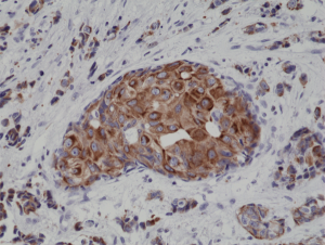
Immunohistochemical staining of formalin fixed and paraffin embedded human breast cancer tissue sections using Anti-CK18 Rabbit Monoclonal Antibody (Clone RM279) at a 1:4000 dilution.
Immunohistochemical staining of formalin fixed and paraffin embedded human breast cancer tissue sections using Anti-CK18 Rabbit Monoclonal Antibody (Clone RM279) at a 1:4000 dilution.
anti-Cytokeratin-18 (human), Rabbit Monoclonal (RM279)
REV-31-1162-00
ApplicationsWestern Blot, ImmunoHistoChemistry
Product group Antibodies
ReactivityHuman
TargetKRT18
Overview
- SupplierRevMAb Biosciences
- Product Nameanti-Cytokeratin-18 (human), Rabbit Monoclonal (RM279)
- Delivery Days Customer2
- ApplicationsWestern Blot, ImmunoHistoChemistry
- CertificationResearch Use Only
- ClonalityMonoclonal
- Clone IDRM279
- Gene ID3875
- Target nameKRT18
- Target descriptionkeratin 18
- Target synonymsCK-18, CYK18, K18, keratin, type I cytoskeletal 18, cell proliferation-inducing gene 46 protein, cytokeratin 18, keratin 18, type I
- HostRabbit
- IsotypeIgG
- Protein IDP05783
- Protein NameKeratin, type I cytoskeletal 18
- Scientific DescriptionCytokeratins are keratin proteins found in the intracytoplasmic cytoskeleton of epithelial tissue (at least 20 different polypeptides). They are an important component of intermediate filaments, which help cells resist mechanical stress. Expression of these cytokeratins within epithelial cells is largely specific to particular organs or tissues. The subsets of cytokeratins which an epithelial cell expresses depends mainly on the type of epithelium, the moment in the course of terminal differentiation and the stage of development. Thus a specific cytokeratin expression profile allows the identification of epithelial cells. Furthermore, this applies also to the malignant counterparts of the epithelia, (carcinomas). Cytokeratin subtype expression patterns are used to an increasing extent in the distinction of different types of epithelial malignancies. The cytokeratin antibodies are not only of assistance in the differential diagnosis of tumors using immunohistochemistry on tissue sections, but are also a useful tool in cytopathology and flow cytometric assays. Cytokeratin-18 (Keratin 18; CK18) belongs to the type A (acidic) subfamily of low molecular weight keratins and exists in combination with cytokeratin-8. Tissues from gastrointestinal tract, respiratory tract and urogenital tract, as well as endocrine and exocrine tissues and mesothelial cells are positive for cytokeratin-18. Further, cytokeratin-18 is found primarily in non-squamous epithelia and is present in a majority of adenocarcinomas and ductal carcinomas, but not in squamous cell carcinomas. Mutations in the cytokeratin-18 gene have been linked to cryptogenic cirrhosis. During apoptosis, keratin filaments are altered and remarkably stable keratin fragments are generated. Activation of caspases leads to proteolysis of numerous cellular proteins, including structural components of epithelial cells. Cytokeratin-18 along with cytokeratin-8 are the major components of intermediate filaments in simple epithelial cells, and the only keratin pair in normal hepatocytes. Cytokeratin-18 is often used to identify differentiated isolated hepatocytes. During apoptosis, cytokeratin-8/cytokeratin-18 fragments are dramatically reorganized and cytokeratin-18 is cleaved by caspases at multiple sequence sites. Cleavage of cytokeratin-18 is an early event occurring during apoptosis. - Recombinant Antibody. This antibody reacts to human CK18 (Cytokeratin-18). Applications: WB, IHC. Source: Rabbit. Liquid. 50% Glycerol/PBS with 1% BSA and 0.09% sodium azide. Cytokeratins are keratin proteins found in the intracytoplasmic cytoskeleton of epithelial tissue (at least 20 different polypeptides). They are an important component of intermediate filaments, which help cells resist mechanical stress. Expression of these cytokeratins within epithelial cells is largely specific to particular organs or tissues. The subsets of cytokeratins which an epithelial cell expresses depends mainly on the type of epithelium, the moment in the course of terminal differentiation and the stage of development. Thus a specific cytokeratin expression profile allows the identification of epithelial cells. Furthermore, this applies also to the malignant counterparts of the epithelia, (carcinomas). Cytokeratin subtype expression patterns are used to an increasing extent in the distinction of different types of epithelial malignancies. The cytokeratin antibodies are not only of assistance in the differential diagnosis of tumors using immunohistochemistry on tissue sections, but are also a useful tool in cytopathology and flow cytometric assays. Cytokeratin-18 (Keratin 18; CK18) belongs to the type A (acidic) subfamily of low molecular weight keratins and exists in combination with cytokeratin-8. Tissues from gastrointestinal tract, respiratory tract and urogenital tract, as well as endocrine and exocrine tissues and mesothelial cells are positive for cytokeratin-18. Further, cytokeratin-18 is found primarily in non-squamous epithelia and is present in a majority of adenocarcinomas and ductal carcinomas, but not in squamous cell carcinomas. Mutations in the cytokeratin-18 gene have been linked to cryptogenic cirrhosis. During apoptosis, keratin filaments are altered and remarkably stable keratin fragments are generated. Activation of caspases leads to proteolysis of numerous cellular proteins, including structural components of epithelial cells. Cytokeratin-18 along with cytokeratin-8 are the major components of intermediate filaments in simple epithelial cells, and the only keratin pair in normal hepatocytes. Cytokeratin-18 is often used to identify differentiated isolated hepatocytes. During apoptosis, cytokeratin-8/cytokeratin-18 fragments are dramatically reorganized and cytokeratin-18 is cleaved by caspases at multiple sequence sites. Cleavage of cytokeratin-18 is an early event occurring during apoptosis.
- ReactivityHuman
- Storage Instruction-20°C,2°C to 8°C
- UNSPSC41116161
Datasheet
Related products
Product group Antibodies
Anti-Keratin 18 AntibodyA93721
ApplicationsImmunoFluorescence, ImmunoPrecipitation, Western Blot, ELISA, ImmunoHistoChemistry
ReactivityHuman, Mouse, Rat
- SizePrice
Product group Antibodies

Anti-Cytokeratin 18 [RGE53]AB03339-1.1
ApplicationsFlow Cytometry, Western Blot, ImmunoHistoChemistry
ReactivityCanine, Chicken, Hamster, Human, Mouse, Porcine, Rabbit, Rat, Zebra Fish
TargetKRT18
- SizePrice

Product group Antibodies
Anti-Cytokeratin 18/KRT18 Antibody Picoband(r)A01357-1-CARRIER-FREE
ApplicationsFlow Cytometry, ImmunoFluorescence, Western Blot, ImmunoCytoChemistry, ImmunoHistoChemistry, ImmunoHistoChemistry Frozen
ReactivityHuman, Mouse, Rat
TargetKRT18
- SizePrice
Product group Antibodies
CK18 Polyclonal Antibodybs-1339R
ApplicationsFlow Cytometry, ImmunoFluorescence, Western Blot, ELISA, ImmunoCytoChemistry, ImmunoHistoChemistry, ImmunoHistoChemistry Frozen, ImmunoHistoChemistry Paraffin
ReactivityCanine, Chicken, Human, Mouse, Rabbit, Rat
TargetKRT18
- SizePrice
Product group Antibodies
KRT18 Monoclonal AntibodyCSB-MA000253
ApplicationsImmunoFluorescence, Western Blot, ELISA, ImmunoHistoChemistry
ReactivityHuman, Mouse, Rat
TargetKRT18
- SizePrice
Product group Antibodies

Goat anti-keratin 18EB12358
ApplicationsWestern Blot, ELISA, ImmunoHistoChemistry
ReactivityBovine, Canine, Human, Mouse, Porcine, Rat
TargetKRT18
- SizePrice
Product group Antibodies
ApplicationsFlow Cytometry
TargetKRT18
- SizePrice

Product group Antibodies
Anti-KRT18 AntibodyHPA001605
ApplicationsWestern Blot, ImmunoCytoChemistry, ImmunoHistoChemistry
ReactivityHuman
TargetKRT18
- SizePrice