


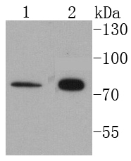
Figure 1. Western blot analysis of Cytokeratin 1/KRT1 using anti-Cytokeratin 1/KRT1 antibody (A01639). Electrophoresis was performed on a 5-20% SDS-PAGE gel at 70V (Stacking gel) / 90V (Resolving gel) for 2-3 hours. The sample well of each lane was loaded with 30 ug of sample under reducing conditions. Lane 1: human A375 whole cell lysates, Lane 2: human Hela whole cell lysates. After electrophoresis, proteins were transferred to a nitrocellulose membrane at 150 mA for 50-90 minutes. Blocked the membrane with 5% non-fat milk/TBS for 1.5 hour at RT. The membrane was incubated with rabbit anti-Cytokeratin 1/KRT1 antigen affinity purified polyclonal antibody (Catalog # A01639) at 0.5 microg/mL overnight at 4°C, then washed with TBS-0.1%Tween 3 times with 5 minutes each and probed with a goat anti-rabbit IgG-HRP secondary antibody at a dilution of 1:5000 for 1.5 hour at RT. The signal is developed using an Enhanced Chemiluminescent detection (ECL) kit (Catalog # EK1002) with Tanon 5200 system. A specific band was detected for Cytokeratin 1/KRT1 at approximately 66 kDa. The expected band size for Cytokeratin 1/KRT1 is at 66 kDa.
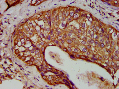
. Cytokeratin 1/KRT1 was detected in a paraffin-embedded section of human skin cancer tissue. Heat mediated antigen retrieval was performed in EDTA buffer (pH 8.0, epitope retrieval solution). The tissue section was blocked with 10% goat serum. The tissue section was then incubated with 2 microg/ml rabbit anti-Cytokeratin 1/KRT1 Antibody (A01639) overnight at 4°C. Peroxidase Conjugated Goat Anti-rabbit IgG was used as secondary antibody and incubated for 30 minutes at 37°C. The tissue section was developed using HRP Conjugated Rabbit IgG Super Vision Assay Kit (Catalog # SV0002) with DAB as the chromogen.")
. Cytokeratin 1/KRT1 was detected in a paraffin-embedded section of human skin cancer tissue. Heat mediated antigen retrieval was performed in EDTA buffer (pH 8.0, epitope retrieval solution). The tissue section was blocked with 10% goat serum. The tissue section was then incubated with 5 microg/mL rabbit anti-Cytokeratin 1/KRT1 Antibody (A01639) overnight at 4°C. DyLight®550 Conjugated Goat Anti-Rabbit IgG (BA1135) was used as secondary antibody at 1:500 dilution and incubated for 30 minutes at 37°C. The section was counterstained with DAPI. Visualize using a fluorescence microscope and filter sets appropriate for the label used.")
. Overlay histogram showing Jurkat cells stained with A01639 (Blue line). To facilitate intracellular staining, cells were fixed with 4% paraformaldehyde and permeabilized with permeabilization buffer. The cells were blocked with 10% normal goat serum. And then incubated with rabbit anti-Cytokeratin 1/KRT1 Antibody (A01639, 1 microg/1x106 cells) for 30 min at 20°C. DyLight®488 conjugated goat anti-rabbit IgG (BA1127, 5-10 microg/1x106 cells) was used as secondary antibody for 30 minutes at 20°C. Isotype control antibody (Green line) was rabbit IgG (1 microg/1x106) used under the same conditions. Unlabelled sample without incubation with primary antibody and secondary antibody (Red line) was used as a blank control.")
Figure 1. Western blot analysis of Cytokeratin 1/KRT1 using anti-Cytokeratin 1/KRT1 antibody (A01639). Electrophoresis was performed on a 5-20% SDS-PAGE gel at 70V (Stacking gel) / 90V (Resolving gel) for 2-3 hours. The sample well of each lane was loaded with 30 ug of sample under reducing conditions. Lane 1: human A375 whole cell lysates, Lane 2: human Hela whole cell lysates. After electrophoresis, proteins were transferred to a nitrocellulose membrane at 150 mA for 50-90 minutes. Blocked the membrane with 5% non-fat milk/TBS for 1.5 hour at RT. The membrane was incubated with rabbit anti-Cytokeratin 1/KRT1 antigen affinity purified polyclonal antibody (Catalog # A01639) at 0.5 microg/mL overnight at 4°C, then washed with TBS-0.1%Tween 3 times with 5 minutes each and probed with a goat anti-rabbit IgG-HRP secondary antibody at a dilution of 1:5000 for 1.5 hour at RT. The signal is developed using an Enhanced Chemiluminescent detection (ECL) kit (Catalog # EK1002) with Tanon 5200 system. A specific band was detected for Cytokeratin 1/KRT1 at approximately 66 kDa. The expected band size for Cytokeratin 1/KRT1 is at 66 kDa.
Anti-Cytokeratin 1/KRT1 Antibody Picoband(r)
A01639-CARRIER-FREE
ApplicationsFlow Cytometry, ImmunoFluorescence, Western Blot, ELISA, ImmunoHistoChemistry
Product group Antibodies
ReactivityHuman
TargetKRT1
Overview
- SupplierBoster Bio
- Product NameAnti-Cytokeratin 1/KRT1 Antibody Picoband(r)
- Delivery Days Customer9
- Application Supplier NoteTested Species: In-house tested species with positive results. Other applications have not been tested. Optimal dilutions should be determined by end users.
- ApplicationsFlow Cytometry, ImmunoFluorescence, Western Blot, ELISA, ImmunoHistoChemistry
- CertificationResearch Use Only
- ClonalityPolyclonal
- Concentration500 ug/ml
- Gene ID3848
- Target nameKRT1
- Target descriptionkeratin 1
- Target synonymsAEI2, CK1, EHK, EHK1, EPPK, K1, KRT1A, NEPPK, keratin, type II cytoskeletal 1, 67 kDa cytokeratin, CK-1, cytokeratin 1, epidermolytic hyperkeratosis 1, hair alpha protein, keratin 1, type II, type-II keratin Kb1
- HostRabbit
- IsotypeIgG
- Protein IDP04264
- Protein NameKeratin, type II cytoskeletal 1
- Scientific DescriptionBoster Bio Anti-Cytokeratin 1/KRT1 Antibody Picoband® catalog # A01639. Tested in ELISA, Flow Cytometry, IF, IHC, WB applications. This antibody reacts with Human. The brand Picoband indicates this is a premium antibody that guarantees superior quality, high affinity, and strong signals with minimal background in Western blot applications. Only our best-performing antibodies are designated as Picoband, ensuring unmatched performance.
- ReactivityHuman
- Storage Instruction-20°C,2°C to 8°C
- UNSPSC12352203
Related products
Product group Antibodies
ApplicationsWestern Blot
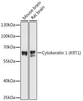
ReactivityMouse, Rat
- SizePrice
Product group Antibodies
Anti-HMW Cytokeratin Antibody188-10212
ReactivityHuman, Mouse, Rat
TargetKRT1
- SizePrice
Product group Antibodies

ApplicationsWestern Blot, ImmunoHistoChemistry
ReactivityBovine, Chicken, Human, Monkey, Mouse, Porcine, Rabbit, Rat
TargetKRT1
- SizePrice
Product group Antibodies

ApplicationsImmunoHistoChemistry
ReactivityHuman
TargetKRT1
- SizePrice
Product group Antibodies
Cytokeratin 1 Recombinant AntibodyBSM-52051R
ApplicationsImmunoFluorescence, Western Blot, ImmunoCytoChemistry, ImmunoHistoChemistry, ImmunoHistoChemistry Frozen, ImmunoHistoChemistry Paraffin
ReactivityHuman, Mouse, Rat
TargetKRT1
- SizePrice
Product group Antibodies
KRT1 AntibodyCSB-PA012503LA01HU
ApplicationsImmunoFluorescence, ELISA, ImmunoHistoChemistry
ReactivityHuman
TargetKRT1
- SizePrice
Product group Antibodies
ApplicationsWestern Blot, ImmunoHistoChemistry
TargetKRT1
- SizePrice
![Non-transfected (–) and transfected (+) 293T whole cell extracts were separated by 7.5% SDS-PAGE, and the membrane was blotted with pan Cytokeratin antibody [HL2253] (GTX638302) diluted at 1:5000. The HRP-conjugated anti-rabbit IgG antibody (GTX213110-01) was used to detect the primary antibody.](https://www.genetex.com/upload/website/prouct_img/normal/GTX638302/GTX638302_T-44960_20240329_WB_multiple_B_24052202_179.webp)
Product group Antibodies
pan Cytokeratin antibody [HL2253]GTX638302
ApplicationsWestern Blot, ImmunoHistoChemistry, ImmunoHistoChemistry Paraffin
ReactivityHuman, Mouse
TargetKRT1
- SizePrice