

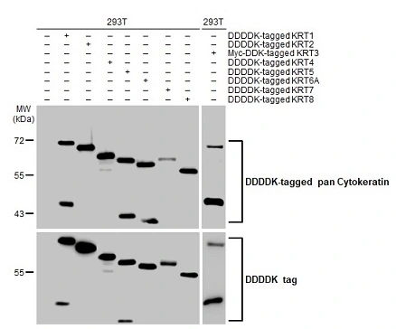
Non-transfected (–) and transfected (+) 293T whole cell extracts were separated by 7.5% SDS-PAGE, and the membrane was blotted with pan Cytokeratin antibody [HL2253] (GTX638302) diluted at 1:5000. The HRP-conjugated anti-rabbit IgG antibody (GTX213110-01) was used to detect the primary antibody.
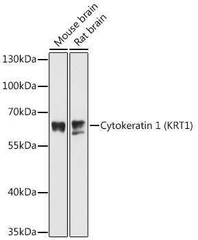
![Whole cell extract (30 μg) was separated by 7.5% SDS-PAGE, and the membrane was blotted with pan Cytokeratin antibody [HL2253] (GTX638302) diluted at 1:1000. The HRP-conjugated anti-rabbit IgG antibody (GTX213110-01) was used to detect the primary antibody.](https://www.genetex.com/upload/website/prouct_img/normal/GTX638302/GTX638302_45425_20240607_WB_24061301_870.webp "Whole cell extract (30 μg) was separated by 7.5% SDS-PAGE, and the membrane was blotted with pan Cytokeratin antibody [HL2253] (GTX638302) diluted at 1:1000. The HRP-conjugated anti-rabbit IgG antibody (GTX213110-01) was used to detect the primary antibody.")
![pan Cytokeratin antibody [HL2253] detects pan Cytokeratin protein by immunohistochemical analysis. Sample: Paraffin-embedded mouse tissues. pan Cytokeratin stained by pan Cytokeratin antibody [HL2253] (GTX638302) diluted at 1:10000. Antigen Retrieval: Tris-EDTA buffer, pH 9.0, 15 min](https://www.genetex.com/upload/website/prouct_img/normal/GTX638302/GTX638302_45425_20250611_IHC-P_M_2_25061901_135.webp "pan Cytokeratin antibody [HL2253] detects pan Cytokeratin protein by immunohistochemical analysis. Sample: Paraffin-embedded mouse tissues. pan Cytokeratin stained by pan Cytokeratin antibody [HL2253] (GTX638302) diluted at 1:10000. Antigen Retrieval: Tris-EDTA buffer, pH 9.0, 15 min")
![pan Cytokeratin antibody [HL2253] detects pan Cytokeratin protein by immunohistochemical analysis. Sample: Paraffin-embedded mouse tissues. pan Cytokeratin stained by pan Cytokeratin antibody [HL2253] (GTX638302) diluted at 1:10000. Antigen Retrieval: Tris-EDTA buffer, pH 9.0, 15 min](https://www.genetex.com/upload/website/prouct_img/normal/GTX638302/GTX638302_45425_20250611_IHC-P_M_1_25061901_929.webp "pan Cytokeratin antibody [HL2253] detects pan Cytokeratin protein by immunohistochemical analysis. Sample: Paraffin-embedded mouse tissues. pan Cytokeratin stained by pan Cytokeratin antibody [HL2253] (GTX638302) diluted at 1:10000. Antigen Retrieval: Tris-EDTA buffer, pH 9.0, 15 min")
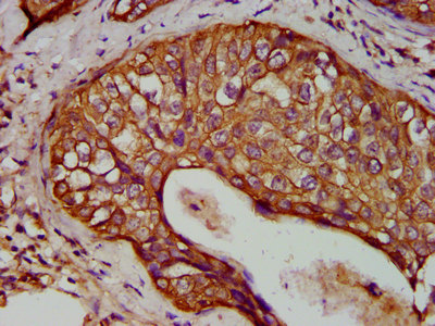
![pan Cytokeratin antibody [HL2253] detects pan Cytokeratin protein by immunohistochemical analysis. Sample: Paraffin-embedded human appendix. pan Cytokeratin stained by pan Cytokeratin antibody [HL2253] (GTX638302) diluted at 1:10000. Antigen Retrieval: Tris-EDTA buffer, pH 9.0, 15 min](https://www.genetex.com/upload/website/prouct_img/normal/GTX638302/GTX638302_45425_20250801_IHC-P_2_25081423_549.webp "pan Cytokeratin antibody [HL2253] detects pan Cytokeratin protein by immunohistochemical analysis. Sample: Paraffin-embedded human appendix. pan Cytokeratin stained by pan Cytokeratin antibody [HL2253] (GTX638302) diluted at 1:10000. Antigen Retrieval: Tris-EDTA buffer, pH 9.0, 15 min")
![pan Cytokeratin antibody [HL2253] detects pan Cytokeratin protein by immunohistochemical analysis. Sample: Paraffin-embedded human tonsil. pan Cytokeratin stained by pan Cytokeratin antibody [HL2253] (GTX638302) diluted at 1:10000. Antigen Retrieval: Tris-EDTA buffer, pH 9.0, 15 min](https://www.genetex.com/upload/website/prouct_img/normal/GTX638302/GTX638302_45425_20250801_IHC-P_1_25081423_817.webp "pan Cytokeratin antibody [HL2253] detects pan Cytokeratin protein by immunohistochemical analysis. Sample: Paraffin-embedded human tonsil. pan Cytokeratin stained by pan Cytokeratin antibody [HL2253] (GTX638302) diluted at 1:10000. Antigen Retrieval: Tris-EDTA buffer, pH 9.0, 15 min")
Non-transfected (–) and transfected (+) 293T whole cell extracts were separated by 7.5% SDS-PAGE, and the membrane was blotted with pan Cytokeratin antibody [HL2253] (GTX638302) diluted at 1:5000. The HRP-conjugated anti-rabbit IgG antibody (GTX213110-01) was used to detect the primary antibody.
pan Cytokeratin antibody [HL2253]
GTX638302
ApplicationsWestern Blot, ImmunoHistoChemistry, ImmunoHistoChemistry Paraffin
Product group Antibodies
ReactivityHuman, Mouse
TargetKRT1
Overview
- SupplierGeneTex
- Product Namepan Cytokeratin antibody [HL2253]
- Delivery Days Customer7
- Application Supplier NoteWB: 1:1000-1:10000. IHC-P: 1:51-1:1000. *Optimal dilutions/concentrations should be determined by the researcher.Not tested in other applications.
- ApplicationsWestern Blot, ImmunoHistoChemistry, ImmunoHistoChemistry Paraffin
- CertificationResearch Use Only
- ClonalityMonoclonal
- Clone IDHL2253
- Concentration1 mg/ml
- ConjugateUnconjugated
- Gene ID3848
- Target nameKRT1
- Target descriptionkeratin 1
- Target synonymsAEI2, CK1, EHK, EHK1, EPPK, K1, KRT1A, NEPPK, keratin, type II cytoskeletal 1, 67 kDa cytokeratin, CK-1, cytokeratin 1, epidermolytic hyperkeratosis 1, hair alpha protein, keratin 1, type II, type-II keratin Kb1
- HostRabbit
- IsotypeIgG
- ReactivityHuman, Mouse
- Storage Instruction-20°C or -80°C,2°C to 8°C
- UNSPSC41116161
Datasheet
Related products
Product group Antibodies
ApplicationsWestern Blot
ReactivityMouse, Rat
- SizePrice
Product group Antibodies
Anti-HMW Cytokeratin Antibody188-10212
ReactivityHuman, Mouse, Rat
TargetKRT1
- SizePrice
Product group Antibodies

ApplicationsWestern Blot, ImmunoHistoChemistry
ReactivityBovine, Chicken, Human, Monkey, Mouse, Porcine, Rabbit, Rat
TargetKRT1
- SizePrice
Product group Antibodies

ApplicationsImmunoHistoChemistry
ReactivityHuman
TargetKRT1
- SizePrice

Product group Antibodies
Anti-Cytokeratin 1/KRT1 Antibody Picoband(r)A01639-CARRIER-FREE
ApplicationsFlow Cytometry, ImmunoFluorescence, Western Blot, ELISA, ImmunoHistoChemistry
ReactivityHuman
TargetKRT1
- SizePrice
Product group Antibodies
Cytokeratin 1 Recombinant AntibodyBSM-52051R
ApplicationsImmunoFluorescence, Western Blot, ImmunoCytoChemistry, ImmunoHistoChemistry, ImmunoHistoChemistry Frozen, ImmunoHistoChemistry Paraffin
ReactivityHuman, Mouse, Rat
TargetKRT1
- SizePrice
Product group Antibodies
KRT1 AntibodyCSB-PA012503LA01HU
ApplicationsImmunoFluorescence, ELISA, ImmunoHistoChemistry
ReactivityHuman
TargetKRT1
- SizePrice
Product group Antibodies
ApplicationsWestern Blot, ImmunoHistoChemistry
TargetKRT1
- SizePrice
Product group Antibodies
pan Cytokeratin antibody [OSCAR]GTX01670
ApplicationsImmunoHistoChemistry, ImmunoHistoChemistry Paraffin
ReactivityHuman
TargetKRT1
- SizePrice