


Immunofluorescent staining of human cell line MCF7 shows localization to nucleus.
Immunofluorescent staining of human cell line MCF7 shows localization to nucleus.
Anti-DCK Antibody
HPA059609
ApplicationsImmunoCytoChemistry
Product group Antibodies
ReactivityHuman
TargetDCK
Overview
- SupplierAtlas Antibodies
- Product NameAnti-DCK Antibody
- Delivery Days Customer4
- ApplicationsImmunoCytoChemistry
- CertificationResearch Use Only
- ClonalityPolyclonal
- ConjugateUnconjugated
- Gene ID1633
- Target nameDCK
- Target descriptiondeoxycytidine kinase
- Target synonymsdeoxycytidine kinase, deoxyadenosine kinase, deoxyguanosine kinase, deoxynucleoside kinase
- HostRabbit
- IsotypeIgG
- Protein IDP27707
- Protein NameDeoxycytidine kinase
- Scientific DescriptionRecombinant Protein Epitope Signature Tag (PrEST) antigen sequence
- ReactivityHuman
- Storage Instruction-20°C,2°C to 8°C
- UNSPSC41116161
Datasheet
MSDS
Related products
Product group Antibodies
Anti-DCK AntibodyA29835
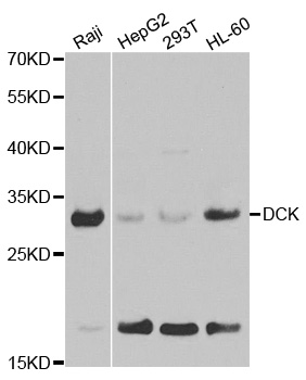
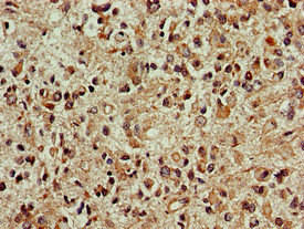
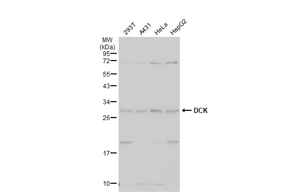
ApplicationsImmunoFluorescence, Western Blot, ImmunoHistoChemistry
ReactivityHuman, Mouse, Rat
- SizePrice
Product group Antibodies
Anti-DCK Antibody144-01794
ApplicationsImmunoFluorescence, Western Blot, ImmunoHistoChemistry
ReactivityHuman, Mouse, Rat
TargetDCK
- SizePrice

Product group Antibodies
Anti-DCK Antibody Picoband(r)A01655-1-CARRIER-FREE
ApplicationsFlow Cytometry, Western Blot, ELISA, ImmunoHistoChemistry
ReactivityHuman, Mouse, Rat
TargetDCK
- SizePrice
Product group Antibodies
DCK Polyclonal AntibodyBS-5749R
ApplicationsImmunoFluorescence, ELISA, ImmunoCytoChemistry, ImmunoHistoChemistry, ImmunoHistoChemistry Frozen, ImmunoHistoChemistry Paraffin
ReactivityEquine, Human, Mouse, Porcine, Rat
TargetDCK
- SizePrice
Product group Antibodies
DCK AntibodyCSB-PA006547LA01HU
ApplicationsELISA, ImmunoHistoChemistry
ReactivityHuman
TargetDCK
- SizePrice
Product group Antibodies

DCK / Deoxycytidine kinase AntibodyLS-C404679
ApplicationsWestern Blot, ELISA, ImmunoHistoChemistry
ReactivityHuman, Mouse, Rat
TargetDCK
- SizePrice
Product group Antibodies
DCK antibodyGTX102800
ApplicationsImmunoFluorescence, ImmunoPrecipitation, Western Blot, ImmunoCytoChemistry, ImmunoHistoChemistry, ImmunoHistoChemistry Paraffin
ReactivityHuman, Mouse, Rat
TargetDCK
- SizePrice

Product group Antibodies
Anti-DCK AntibodyHPA062773
ApplicationsWestern Blot, ImmunoHistoChemistry
ReactivityHuman
TargetDCK
- SizePrice