

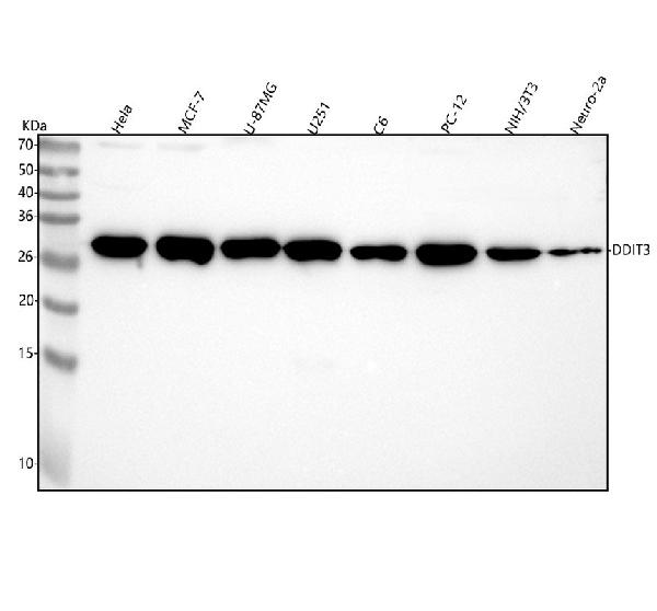
Figure 1. Western blot analysis of DDIT3 using anti-DDIT3 antibody (M00311). Electrophoresis was performed on a 5-20% SDS-PAGE gel at 70V (Stacking gel) / 90V (Resolving gel) for 2-3 hours. The sample well of each lane was loaded with 30 ug of sample under reducing conditions. Lane 1: human Hela whole cell lysates, Lane 2: human MCF-7 whole cell lysates, Lane 3: human U-87MG whole cell lysates, Lane 4: human U251 whole cell lysates, Lane 5: rat C6 whole cell lysates, Lane 6: rat PC-12 whole cell lysates, Lane 7: mouse NIH/3T3 whole cell lysates, Lane 8: mouse Neuro-2a whole cell lysates. After electrophoresis, proteins were transferred to a nitrocellulose membrane at 150 mA for 50-90 minutes. Blocked the membrane with 5% non-fat milk/TBS for 1.5 hour at RT. The membrane was incubated with rabbit anti-DDIT3 antigen affinity purified monoclonal antibody (Catalog # M00311) at 1:500 overnight at 4°C, then washed with TBS-0.1%Tween 3 times with 5 minutes each and probed with a goat anti-rabbit IgG-HRP secondary antibody at a dilution of 1:500 for 1.5 hour at RT. The signal is developed using an Enhanced Chemiluminescent detection (ECL) kit (Catalog # EK1002) with Tanon 5200 system. A specific band was detected for DDIT3 at approximately 29 kDa. The expected band size for DDIT3 is at 20 kDa.
Figure 1. Western blot analysis of DDIT3 using anti-DDIT3 antibody (M00311). Electrophoresis was performed on a 5-20% SDS-PAGE gel at 70V (Stacking gel) / 90V (Resolving gel) for 2-3 hours. The sample well of each lane was loaded with 30 ug of sample under reducing conditions. Lane 1: human Hela whole cell lysates, Lane 2: human MCF-7 whole cell lysates, Lane 3: human U-87MG whole cell lysates, Lane 4: human U251 whole cell lysates, Lane 5: rat C6 whole cell lysates, Lane 6: rat PC-12 whole cell lysates, Lane 7: mouse NIH/3T3 whole cell lysates, Lane 8: mouse Neuro-2a whole cell lysates. After electrophoresis, proteins were transferred to a nitrocellulose membrane at 150 mA for 50-90 minutes. Blocked the membrane with 5% non-fat milk/TBS for 1.5 hour at RT. The membrane was incubated with rabbit anti-DDIT3 antigen affinity purified monoclonal antibody (Catalog # M00311) at 1:500 overnight at 4°C, then washed with TBS-0.1%Tween 3 times with 5 minutes each and probed with a goat anti-rabbit IgG-HRP secondary antibody at a dilution of 1:500 for 1.5 hour at RT. The signal is developed using an Enhanced Chemiluminescent detection (ECL) kit (Catalog # EK1002) with Tanon 5200 system. A specific band was detected for DDIT3 at approximately 29 kDa. The expected band size for DDIT3 is at 20 kDa.
Anti-DDIT3/Chop Rabbit Monoclonal Antibody
M00311
ApplicationsFlow Cytometry, Western Blot, ImmunoHistoChemistry
Product group Antibodies
ReactivityHuman, Mouse, Rat
TargetDDIT3
Overview
- SupplierBoster Bio
- Product NameAnti-DDIT3/Chop Rabbit Monoclonal Antibody
- Delivery Days Customer9
- ApplicationsFlow Cytometry, Western Blot, ImmunoHistoChemistry
- CertificationResearch Use Only
- ClonalityMonoclonal
- Clone IDAOHF-4
- Gene ID1649
- Target nameDDIT3
- Target descriptionDNA damage inducible transcript 3
- Target synonymsAltDDIT3, C/EBPzeta, CEBPZ, CHOP, CHOP-10, CHOP10, GADD153, DNA damage-inducible transcript 3 protein, C/EBP zeta, CCAAT/enhancer-binding protein homologous protein, alternative DDIT3 protein, c/EBP-homologous protein 10, growth arrest and DNA damage-inducible protein GADD153
- HostRabbit
- IsotypeIgG
- Protein IDP35638
- Protein NameDNA damage-inducible transcript 3 protein
- Scientific DescriptionBoster Bio Anti-DDIT3/Chop Rabbit Monoclonal Antibody catalog # M00311. Tested in WB, IHC, Flow Cytometry applications. This antibody reacts with Human, Mouse, Rat.
- ReactivityHuman, Mouse, Rat
- Storage Instruction-20°C
- UNSPSC12352203
References
- Sui Y, Feng X, Ma Y, et al. BHBA attenuates endoplasmic reticulum stress-dependent neuroinflammation via the gut-brain axis in a mouse model of heat stress. CNS Neurosci Ther. 2024,30(7):e14840. doi: 10.1111/cns.14840Read this paper
- Xiang X, Xu M, Liu L, et al. Liproxstatin-1 attenuates acute hypertriglyceridemic pancreatitis through inhibiting ferroptosis in rats. Sci Rep. 2024,14(1):9548. doi: 10.1038/s41598-024-60159-7Read this paper
- Li C, Ma Y, Chai X, et al. Ketogenic diet attenuates cognitive dysfunctions induced by hypoglycemia via inhibiting endoplasmic reticulum stress-dependent pathways. Food Funct. 2024,15(3):1294-1309. doi: 10.1039/d3fo04007kRead this paper
- Caglayan M, Ozden S. Potential impacts of bisphenols on prostate cells: An overview of cytotoxicity, proliferation, oxidative stress, apoptosis, and ER-stress response activation. Food Chem Toxicol. 2024,184:114416. doi: 10.1016/j.fct.2023.114416Read this paper
- Izadi MS, Eskandari F, Binayi F, et al. Oxidative and endoplasmic reticulum stress develop adverse metabolic effects due to the high-fat high-fructose diet consumption from birth to young adulthood. Life Sci. 2022,309:120924. doi: 10.1016/j.lfs.2022.120924Read this paper
- Guo Y, Yang C, Guo R, et al. CHOP Regulates Endoplasmic Reticulum Stress-Mediated Hepatoxicity Induced by Monocrotaline. Front Pharmacol. 2021,12:685895. doi: 10.3389/fphar.2021.685895Read this paper
- Binayi F, Zardooz H, Ghasemi R, et al. The chemical chaperon 4-phenyl butyric acid restored high-fat diet- induced hippocampal insulin content and insulin receptor level reduction along with spatial learning and memory deficits in male rats. Physiol Behav. 2021,231:113312. doi: 10.1016/j.physbeh.2021.113312Read this paper
- Wu C, Yuan G, Mo R, et al. Effect of endoplasmic reticulum stress involved in manganese‑induced neurotoxicity in rats. Mol Med Rep. 2019,19(6):5169-5176. doi: 10.3892/mmr.2019.10175Read this paper
- Li X, Wang X, Wang Y, et al. Inhibition of transient receptor potential melastatin 7 (TRPM7) channel induces RA FLSs apoptosis through endoplasmic reticulum (ER) stress. Clin Rheumatol. 2014,33(11):1565-74. doi: 10.1007/s10067-014-2599-xRead this paper
- Cao Y, Hao Y, Li H, et al. Role of endoplasmic reticulum stress in apoptosis of differentiated mouse podocytes induced by high glucose. Int J Mol Med. 2014,33(4):809-16. doi: 10.3892/ijmm.2014.1642Read this paper
Datasheet
MSDS
Related products
Product group Antibodies
Ddit3 Polyclonal AntibodyCAC07500
ApplicationsImmunoFluorescence, ELISA, ImmunoHistoChemistry
TargetDDIT3
- SizePrice
Product group Antibodies
References
DDIT3 Polyclonal AntibodyBS-1361R
ApplicationsFlow Cytometry, ImmunoFluorescence, Western Blot, ELISA, ImmunoCytoChemistry, ImmunoHistoChemistry, ImmunoHistoChemistry Frozen, ImmunoHistoChemistry Paraffin
ReactivityHuman, Mouse, Rat
TargetDDIT3
- SizePrice
Product group Antibodies
Anti-DDIT3 / CHOP Antibody144-61577
ApplicationsImmunoFluorescence, Western Blot, ImmunoHistoChemistry
ReactivityHuman, Mouse, Rat
TargetDDIT3
- SizePrice
Product group Antibodies
Anti-GADD153 AntibodyA94972
ApplicationsImmunoFluorescence, Western Blot, ELISA, ImmunoHistoChemistry
ReactivityHuman, Mouse, Rat
- SizePrice
Product group Antibodies
GADD153 antibodyGTX109226
ApplicationsImmunoFluorescence, Western Blot, ImmunoCytoChemistry
ReactivityHuman, Mouse
TargetDDIT3
- SizePrice
Product group Antibodies
anti-DDIT3 (human), Rabbit Monoclonal (RM485)REV-31-1377-00
ApplicationsWestern Blot
ReactivityHuman
TargetDDIT3
- SizePrice
Product group Antibodies

DDIT3 / CHOP Antibody (clone 1E1)LS-C765520
ApplicationsImmunoHistoChemistry, ImmunoHistoChemistry Paraffin
ReactivityHuman, Mouse, Rat
TargetDDIT3
- SizePrice