


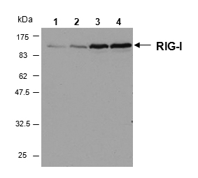
Figure 1. Western blot analysis of DDX58 using anti-DDX58 antibody (A00244-2). Electrophoresis was performed on a 5-20% SDS-PAGE gel at 70V (Stacking gel) / 90V (Resolving gel) for 2-3 hours. The sample well of each lane was loaded with 30 ug of sample under reducing conditions. Lane 1: human HepG2 whole cell lysates, Lane 2: rat C6 whole cell lysates, Lane 3: human Hela whole cell lysates, Lane 4: mouse RAW264.7 whole cell lysates. After electrophoresis, proteins were transferred to a nitrocellulose membrane at 150 mA for 50-90 minutes. Blocked the membrane with 5% non-fat milk/TBS for 1.5 hour at RT. The membrane was incubated with rabbit anti-DDX58 antigen affinity purified polyclonal antibody (Catalog # A00244-2) at 0.5 microg/mL overnight at 4°C, then washed with TBS-0.1%Tween 3 times with 5 minutes each and probed with a goat anti-rabbit IgG-HRP secondary antibody at a dilution of 1:5000 for 1.5 hour at RT. The signal is developed using an Enhanced Chemiluminescent detection (ECL) kit (Catalog # EK1002) with Tanon 5200 system. A specific band was detected for DDX58 at approximately 107 kDa. The expected band size for DDX58 is at 107 kDa.
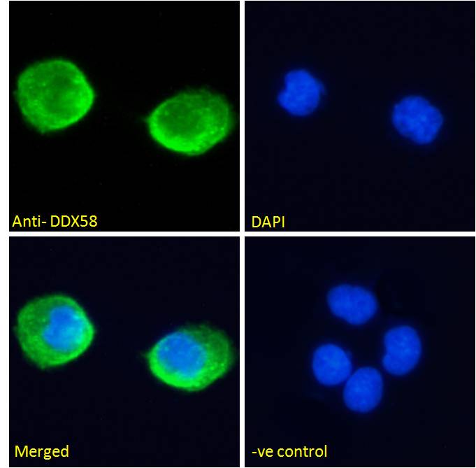
. DDX58 was detected in immunocytochemical section of U20S cells. Enzyme antigen retrieval was performed using IHC enzyme antigen retrieval reagent (AR0022) for 15 mins. The cells were blocked with 10% goat serum. And then incubated with 2microg/mL rabbit anti-DDX58 Antibody (A00244-2) overnight at 4°C. DyLight®594 Conjugated Goat Anti-Rabbit IgG (BA1142) was used as secondary antibody at 1:100 dilution and incubated for 30 minutes at 37°C. The section was counterstained with DAPI. Visualize using a fluorescence microscope and filter sets appropriate for the label used.")
. DDX58 was detected in a paraffin-embedded section of human intestine cancer tissue. Heat mediated antigen retrieval was performed in EDTA buffer (pH 8.0, epitope retrieval solution). The tissue section was blocked with 10% goat serum. The tissue section was then incubated with 5 microg/mL rabbit anti-DDX58 Antibody (A00244-2) overnight at 4°C. DyLight®550 Conjugated Goat Anti-Rabbit IgG (BA1135) was used as secondary antibody at 1:500 dilution and incubated for 30 minutes at 37°C. The section was counterstained with DAPI. Visualize using a fluorescence microscope and filter sets appropriate for the label used.")
. Overlay histogram showing A431 cells stained with A00244-2 (Blue line). To facilitate intracellular staining, cells were fixed with 4% paraformaldehyde and permeabilized with permeabilization buffer. The cells were blocked with 10% normal goat serum. And then incubated with rabbit anti-DDX58 Antibody (A00244-2,1microg/1x106 cells) for 30 min at 20°C. DyLight®488 conjugated goat anti-rabbit IgG (BA1127, 5-10microg/1x106 cells) was used as secondary antibody for 30 minutes at 20°C. Isotype control antibody (Green line) was rabbit IgG (1microg/1x106) used under the same conditions. Unlabelled sample without incubation with primary antibody and secondary antibody (Red line) was used as a blank control.")
Figure 1. Western blot analysis of DDX58 using anti-DDX58 antibody (A00244-2). Electrophoresis was performed on a 5-20% SDS-PAGE gel at 70V (Stacking gel) / 90V (Resolving gel) for 2-3 hours. The sample well of each lane was loaded with 30 ug of sample under reducing conditions. Lane 1: human HepG2 whole cell lysates, Lane 2: rat C6 whole cell lysates, Lane 3: human Hela whole cell lysates, Lane 4: mouse RAW264.7 whole cell lysates. After electrophoresis, proteins were transferred to a nitrocellulose membrane at 150 mA for 50-90 minutes. Blocked the membrane with 5% non-fat milk/TBS for 1.5 hour at RT. The membrane was incubated with rabbit anti-DDX58 antigen affinity purified polyclonal antibody (Catalog # A00244-2) at 0.5 microg/mL overnight at 4°C, then washed with TBS-0.1%Tween 3 times with 5 minutes each and probed with a goat anti-rabbit IgG-HRP secondary antibody at a dilution of 1:5000 for 1.5 hour at RT. The signal is developed using an Enhanced Chemiluminescent detection (ECL) kit (Catalog # EK1002) with Tanon 5200 system. A specific band was detected for DDX58 at approximately 107 kDa. The expected band size for DDX58 is at 107 kDa.
Anti-DDX58 Antibody Picoband(r)
A00244-2-CARRIER-FREE
ApplicationsFlow Cytometry, ImmunoFluorescence, Western Blot, ImmunoCytoChemistry
Product group Antibodies
ReactivityHuman, Mouse, Rat
TargetRIGI
Overview
- SupplierBoster Bio
- Product NameAnti-DDX58 Antibody Picoband(r)
- Delivery Days Customer9
- ApplicationsFlow Cytometry, ImmunoFluorescence, Western Blot, ImmunoCytoChemistry
- CertificationResearch Use Only
- ClonalityPolyclonal
- Concentration500 ug/ml
- Gene ID23586
- Target nameRIGI
- Target descriptionRNA sensor RIG-I
- Target synonymsDDX58, RIG-I, RIG1, RLR-1, SGMRT2, antiviral innate immune response receptor RIG-I, ATP-dependent RNA helicase DDX58, DEAD (Asp-Glu-Ala-Asp) box polypeptide 58, DEAD box protein 58, DEAD/H (Asp-Glu-Ala-Asp/His) box polypeptide, DExD/H-box helicase 58, RNA helicase RIG-I, probable ATP-dependent RNA helicase DDX58, retinoic acid-inducible gene 1 protein, retinoic acid-inducible gene I protein
- HostRabbit
- IsotypeIgG
- Protein IDO95786
- Protein NameAntiviral innate immune response receptor RIG-I
- Scientific DescriptionBoster Bio Anti-DDX58 Antibody Picoband® catalog # A00244-2. Tested in Flow Cytometry, IF, ICC, WB applications. This antibody reacts with Human, Mouse, Rat. The brand Picoband indicates this is a premium antibody that guarantees superior quality, high affinity, and strong signals with minimal background in Western blot applications. Only our best-performing antibodies are designated as Picoband, ensuring unmatched performance.
- ReactivityHuman, Mouse, Rat
- Storage Instruction-20°C,2°C to 8°C
- UNSPSC12352203
Related products
Product group Antibodies
Anti-RIG-I/DDX58 AntibodyA285978
ApplicationsFlow Cytometry, ImmunoFluorescence, ELISA, ImmunoHistoChemistry
ReactivityHuman
- SizePrice
Product group Antibodies
anti-RIG-I, mAb (Alme-1)AG-20B-0009
ApplicationsImmunoPrecipitation, Western Blot, ImmunoHistoChemistry
ReactivityHuman, Mouse
TargetRIGI
- SizePrice
Product group Antibodies
Anti-DDX58 Antibody144-00550
ApplicationsImmunoFluorescence, ImmunoPrecipitation, Western Blot, ImmunoHistoChemistry
ReactivityHuman
TargetRIGI
- SizePrice
Product group Antibodies

DDX58 / RIG-1 / RIG-I AntibodyLS-C831077
ApplicationsELISA, ImmunoHistoChemistry
ReactivityHuman
TargetRIGI
- SizePrice
Product group Antibodies
DDX58 Polyclonal AntibodyBS-0993R
ApplicationsWestern Blot, ELISA, ImmunoHistoChemistry, ImmunoHistoChemistry Paraffin
ReactivityBovine, Canine, Human, Mouse, Porcine, Rat
TargetRIGI
- SizePrice
Product group Antibodies
DDX58 AntibodyCSB-PA006638LA01HU
ApplicationsELISA
ReactivityHuman
TargetRIGI
- SizePrice
Product group Antibodies

Goat anti-DDX58EB08003
ApplicationsFlow Cytometry, ImmunoFluorescence, ELISA, ImmunoHistoChemistry
ReactivityHuman, Mouse
TargetRIGI
- SizePrice
Product group Antibodies
ApplicationsImmunoPrecipitation, Western Blot, ImmunoCytoChemistry, ImmunoHistoChemistry
ReactivityMouse, Rat
TargetRIGI
- SizePrice
Product group Antibodies
DDX58 antibodyGTX132517
ApplicationsWestern Blot
ReactivityHuman
TargetRIGI
- SizePrice