


Immunofluorescent staining of human cell line U2OS shows localization to cytosol.
Immunofluorescent staining of human cell line U2OS shows localization to cytosol.
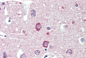
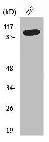
Anti-DLGAP1 Antibody
HPA078234
ApplicationsImmunoCytoChemistry
Product group Antibodies
ReactivityHuman
TargetDLGAP1
Overview
- SupplierAtlas Antibodies
- Product NameAnti-DLGAP1 Antibody
- Delivery Days Customer12
- ApplicationsImmunoCytoChemistry
- CertificationResearch Use Only
- ClonalityPolyclonal
- ConjugateUnconjugated
- Gene ID9229
- Target nameDLGAP1
- Target descriptionDLG associated protein 1
- Target synonymsDAP-1, DAP-1-ALPHA, DAP-1-BETA, DAP1, DLGAP1A, DLGAP1B, GKAP, SAPAP1, disks large-associated protein 1, PSD-95/SAP90 binding protein 1, SAP90/PSD-95-associated protein 1, discs large homolog associated protein 1, guanylate kinase-associated protein
- HostRabbit
- IsotypeIgG
- Protein IDO14490
- Protein NameDisks large-associated protein 1
- Scientific DescriptionRecombinant Protein Epitope Signature Tag (PrEST) antigen sequence
- ReactivityHuman
- Storage Instruction-20°C,2°C to 8°C
- UNSPSC41116161
MSDS
Related products

Product group Antibodies
Anti-DLGAP1 Antibody Picoband(r)A08230-2-CARRIER-FREE
ApplicationsFlow Cytometry, ImmunoFluorescence, Western Blot, ELISA, ImmunoCytoChemistry, ImmunoHistoChemistry
ReactivityHuman, Mouse, Rat
TargetDLGAP1
- SizePrice
Product group Antibodies
Anti-DLGAP1 AntibodyA326240
ApplicationsELISA, ImmunoHistoChemistry
ReactivityHuman
- SizePrice
Product group Antibodies
DLGAP1 AntibodyCSB-PA004048
ApplicationsImmunoFluorescence, Western Blot, ELISA, ImmunoHistoChemistry
ReactivityHuman, Mouse, Rat
TargetDLGAP1
- SizePrice
Product group Antibodies

Goat anti-DLGAP1 / GKAPEB09176
ApplicationsELISA, ImmunoHistoChemistry
ReactivityHuman
TargetDLGAP1
- SizePrice
Product group Antibodies

DLGAP1 Antibody (C-Terminus)LS-C368807
ApplicationsWestern Blot
ReactivityCanine, Chicken, Human, Mouse, Rat
TargetDLGAP1
- SizePrice
![WB analysis of HEK293 (1) and DLGAP1(AA: 490-663)-hIgGFc transfected HEK293 (2) cell lysate using GTX60469 DLGAP1 antibody [3G4].](https://www.genetex.com/upload/website/prouct_img/normal/GTX60469/GTX60469_20170912_WB_w_23061123_985.webp)
Product group Antibodies
DLGAP1 antibody [3G4]GTX60469
ApplicationsImmunoFluorescence, Western Blot, ELISA, ImmunoCytoChemistry, ImmunoHistoChemistry, ImmunoHistoChemistry Paraffin
ReactivityHuman, Mouse
TargetDLGAP1
- SizePrice