


Anti-DRD5 Antibody
A285965
ApplicationsFlow Cytometry, ImmunoFluorescence, ELISA
Product group Antibodies
ReactivityHuman
Overview
- SupplierAntibodies.com
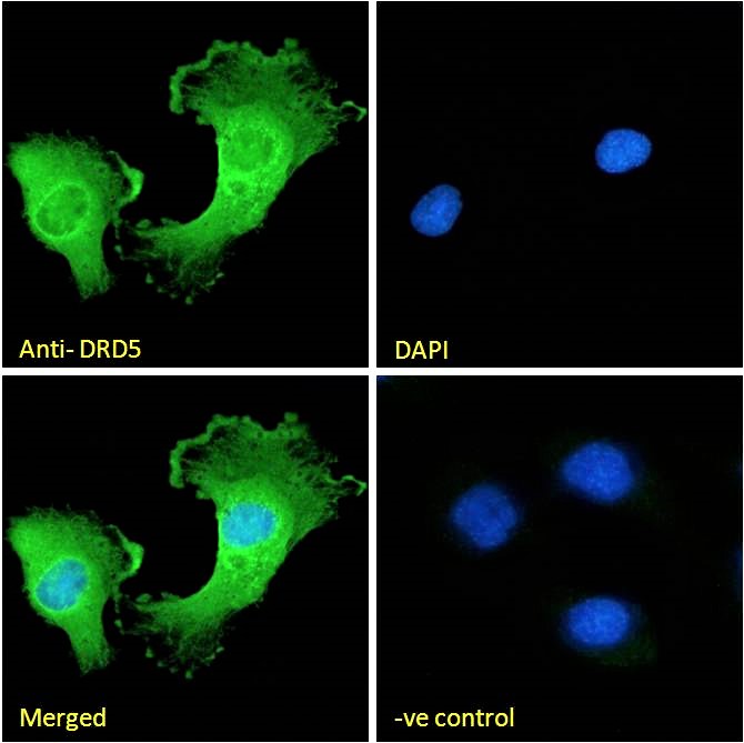
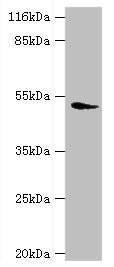
- Product NameAnti-DRD5 Antibody
- Delivery Days Customer7
- ApplicationsFlow Cytometry, ImmunoFluorescence, ELISA
- CertificationResearch Use Only
- ClonalityPolyclonal
- Concentration500 ug/ml
- ConjugateUnconjugated
- HostGoat
- IsotypeIgG
- Scientific DescriptionGoat polyclonal antibody to DRD5.
- ReactivityHuman
- UNSPSC12352203
Related products

Product group Antibodies
Anti-DRD5 Antibody Picoband(r)A03010-1-CARRIER-FREE
ApplicationsFlow Cytometry, Western Blot, ELISA
ReactivityHuman, Mouse, Rat
TargetDRD5
- SizePrice
Product group Antibodies
Anti-PDP2 Antibody144-64831
ApplicationsImmunoFluorescence, Western Blot
ReactivityHuman, Mouse, Rat
TargetDRD5
- SizePrice
Product group Antibodies
DRD5 AntibodyCSB-PA007182EA01HU
ApplicationsImmunoFluorescence, Western Blot, ELISA, ImmunoHistoChemistry
ReactivityHuman, Mouse
TargetDRD5
- SizePrice
Product group Antibodies

Goat anti-DRD5EB07424
ApplicationsFlow Cytometry, ImmunoFluorescence, ELISA
ReactivityHuman
TargetDRD5
- SizePrice
Product group Antibodies
Drd5 Polyclonal AntibodyCAC07349
ApplicationsImmunoFluorescence, Western Blot, ELISA, ImmunoHistoChemistry
ReactivityMouse
TargetDRD5
- SizePrice
Product group Antibodies

DRD5 / Dopamine Receptor D5 AntibodyLS-C400700
ApplicationsWestern Blot, ELISA, ImmunoHistoChemistry
ReactivityHuman
TargetDRD5
- SizePrice
![Dopamine Receptor D5 antibody [HL3081] detects Dopamine Receptor D5 protein by immunofluorescent analysis. Sample: SH-SY-5Y cells were fixed in ice-cold MeOH for 5 min. Green: Dopamine Receptor D5 stained by Dopamine Receptor D5 antibody [HL3081] (GTX640527) diluted at 1:500. Red: alpha Tubulin, a cytoskeleton marker, stained by alpha Tubulin antibody [GT114] (GTX628802) diluted at 1:1000. Blue: Fluoroshield with DAPI (GTX30920).](https://www.genetex.com/upload/website/prouct_img/normal/GTX640527/GTX640527_T-45446_20240712_ICC_IF_24080622_450.webp)
Product group Antibodies
ApplicationsImmunoFluorescence, ImmunoCytoChemistry, ImmunoHistoChemistry, ImmunoHistoChemistry Paraffin
ReactivityHuman, Mouse
TargetDRD5
- SizePrice
Product group Antibodies
Anti-PDP2 AntibodyCAB17190
ApplicationsImmunoFluorescence, Western Blot, ELISA, ImmunoCytoChemistry
ReactivityMouse
TargetDRD5
- SizePrice