


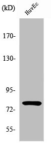
Figure 1. Western blot analysis of PIAS1 using anti-PIAS1 antibody (A01707). Electrophoresis was performed on a 5-20% SDS-PAGE gel at 70V (Stacking gel) / 90V (Resolving gel) for 2-3 hours. The sample well of each lane was loaded with 50ug of sample under reducing conditions. Lane 1: human HepG2 whole cell lysates, Lane 2: human Jurkat whole cell lysates, Lane 3: human HEK293 whole cell lysates, Lane 4: human Raji whole cell lysates, Lane 5: human K562 whole cell lysates, Lane 6: human Hela whole cell lysates. After Electrophoresis, proteins were transferred to a Nitrocellulose membrane at 150mA for 50-90 minutes. Blocked the membrane with 5% Non-fat Milk/ TBS for 1.5 hour at RT. The membrane was incubated with rabbit anti-PIAS1 antigen affinity purified polyclonal antibody (Catalog # A01707) at 0.5 microg/mL overnight at 4°C, then washed with TBS-0.1%Tween 3 times with 5 minutes each and probed with a goat anti-rabbit IgG-HRP secondary antibody at a dilution of 1:5000 for 1.5 hour at RT. The signal is developed using an Enhanced Chemiluminescent detection (ECL) kit (Catalog # EK1002) with Tanon 5200 system. A specific band was detected for PIAS1 at approximately 72KD. The expected band size for PIAS1 is at 72KD.
. PIAS1 was detected in immunocytochemical section of A431 cells. Enzyme antigen retrieval was performed using IHC enzyme antigen retrieval reagent (AR0022) for 15 mins. The cells were blocked with 10% goat serum. And then incubated with 2microg/mL rabbit anti-PIAS1 Antibody (A01707) overnight at 4°C. DyLight®488 Conjugated Goat Anti-Rabbit IgG (BA1127) was used as secondary antibody at 1:100 dilution and incubated for 30 minutes at 37°C. The section was counterstained with DAPI. Visualize using a fluorescence microscope and filter sets appropriate for the label used.")
. Overlay histogram showing PC-3 cells stained with A01707 (Blue line). To facilitate intracellular staining, cells were fixed with 4% paraformaldehyde and permeabilized with permeabilization buffer. The cells were blocked with 10% normal goat serum. And then incubated with rabbit anti-PIAS1 Antibody (A01707, 1microg/1x106 cells) for 30 min at 20°C. DyLight®488 conjugated goat anti-rabbit IgG (BA1127, 5-10microg/1x106 cells) was used as secondary antibody for 30 minutes at 20°C. Isotype control antibody (Green line) was rabbit IgG (1microg/1x106) used under the same conditions. Unlabelled sample without incubation with primary antibody and secondary antibody (Red line) was used as a blank control.")
Figure 1. Western blot analysis of PIAS1 using anti-PIAS1 antibody (A01707). Electrophoresis was performed on a 5-20% SDS-PAGE gel at 70V (Stacking gel) / 90V (Resolving gel) for 2-3 hours. The sample well of each lane was loaded with 50ug of sample under reducing conditions. Lane 1: human HepG2 whole cell lysates, Lane 2: human Jurkat whole cell lysates, Lane 3: human HEK293 whole cell lysates, Lane 4: human Raji whole cell lysates, Lane 5: human K562 whole cell lysates, Lane 6: human Hela whole cell lysates. After Electrophoresis, proteins were transferred to a Nitrocellulose membrane at 150mA for 50-90 minutes. Blocked the membrane with 5% Non-fat Milk/ TBS for 1.5 hour at RT. The membrane was incubated with rabbit anti-PIAS1 antigen affinity purified polyclonal antibody (Catalog # A01707) at 0.5 microg/mL overnight at 4°C, then washed with TBS-0.1%Tween 3 times with 5 minutes each and probed with a goat anti-rabbit IgG-HRP secondary antibody at a dilution of 1:5000 for 1.5 hour at RT. The signal is developed using an Enhanced Chemiluminescent detection (ECL) kit (Catalog # EK1002) with Tanon 5200 system. A specific band was detected for PIAS1 at approximately 72KD. The expected band size for PIAS1 is at 72KD.
Anti-E3 SUMO-protein ligase PIAS1 PIAS1 Antibody Picoband(r)
A01707-CARRIER-FREE
ApplicationsFlow Cytometry, ImmunoFluorescence, Western Blot, ELISA, ImmunoCytoChemistry
Product group Antibodies
ReactivityHuman
TargetPIAS1
Overview
- SupplierBoster Bio
- Product NameAnti-E3 SUMO-protein ligase PIAS1 PIAS1 Antibody Picoband(r)
- Delivery Days Customer9
- Application Supplier NoteTested Species: In-house tested species with positive results. Other applications have not been tested. Optimal dilutions should be determined by end users.
- ApplicationsFlow Cytometry, ImmunoFluorescence, Western Blot, ELISA, ImmunoCytoChemistry
- CertificationResearch Use Only
- ClonalityPolyclonal
- Concentration500 ug/ml
- Gene ID8554
- Target namePIAS1
- Target descriptionprotein inhibitor of activated STAT 1
- Target synonymsDDXBP1, GBP, GU/RH-II, ZMIZ3, E3 SUMO-protein ligase PIAS1, AR interacting protein, DEAD/H (Asp-Glu-Ala-Asp/His) box binding protein 1, DEAD/H box-binding protein 1, E3 SUMO-protein transferase PIAS1, RNA helicase II-binding protein, gu-binding protein, protein inhibitor of activated STAT protein 1, zinc finger, MIZ-type containing 3
- HostRabbit
- IsotypeIgG
- Protein IDO75925
- Protein NameE3 SUMO-protein ligase PIAS1
- Scientific DescriptionBoster Bio Anti-E3 SUMO-protein ligase PIAS1 PIAS1 Antibody catalog # A01707. Tested in ELISA, Flow Cytometry, IF, ICC, WB applications. This antibody reacts with Human. The brand Picoband indicates this is a premium antibody that guarantees superior quality, high affinity, and strong signals with minimal background in Western blot applications. Only our best-performing antibodies are designated as Picoband, ensuring unmatched performance.
- ReactivityHuman
- Storage Instruction-20°C,2°C to 8°C
- UNSPSC12352203
Related products
Product group Antibodies
Anti-PIAS1 AntibodyA97348
ApplicationsWestern Blot, ELISA, ImmunoHistoChemistry
ReactivityHuman, Mouse
- SizePrice
Product group Antibodies
Anti-PIAS1 Antibody144-61705
ApplicationsWestern Blot
ReactivityHuman, Mouse
TargetPIAS1
- SizePrice
Product group Antibodies
PIAS1/2 Recombinant AntibodyBSM-62931R
ApplicationsFlow Cytometry, ImmunoFluorescence, ImmunoPrecipitation, Western Blot, ImmunoCytoChemistry, ImmunoHistoChemistry, ImmunoHistoChemistry Frozen, ImmunoHistoChemistry Paraffin
ReactivityHuman, Mouse, Rat
TargetPIAS1
- SizePrice
Product group Antibodies
PIAS1 AntibodyCSB-PA003776
ApplicationsWestern Blot, ELISA, ImmunoHistoChemistry
ReactivityHuman, Mouse
TargetPIAS1
- SizePrice
Product group Antibodies
ApplicationsImmunoPrecipitation, Western Blot, ImmunoCytoChemistry, ImmunoHistoChemistry
TargetPIAS1
- SizePrice
Product group Antibodies
PIAS1 antibodyGTX31410
ApplicationsWestern Blot, ELISA, ImmunoHistoChemistry, ImmunoHistoChemistry Paraffin
ReactivityHuman, Mouse, Rat
TargetPIAS1
- SizePrice
Product group Antibodies

PIAS1 AntibodyLS-C346096
ApplicationsImmunoFluorescence, Western Blot, ImmunoHistoChemistry
ReactivityHuman, Mouse
TargetPIAS1
- SizePrice