


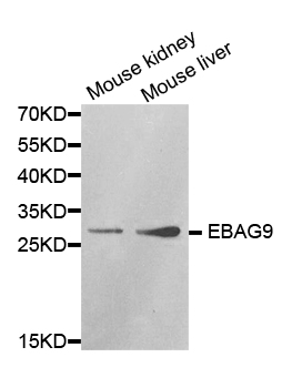
Figure 1. Western blot analysis of EBAG9 using anti-EBAG9 antibody (PB9553). Electrophoresis was performed on a 5-20% SDS-PAGE gel at 70V (Stacking gel) / 90V (Resolving gel) for 2-3 hours. Lane 1: Rat Testis Tissue Lysate at 50ug, Lane 2: 22RV1 Whole Cell Lysate at 40ug, Lane 3: HELA Whole Cell Lysate at 40ug, Lane 4: MCF-7 Whole Cell Lysate at 40ug, Lane 5: JURKAT Whole Cell Lysate at 40ug. After electrophoresis, proteins were transferred to a nitrocellulose membrane at 150 mA for 50-90 minutes. Blocked the membrane with 5% non-fat milk/TBS for 1.5 hour at RT. The membrane was incubated with rabbit anti-EBAG9 antigen affinity purified polyclonal antibody (Catalog # PB9553) at 0.5 microg/mL overnight at 4°C, then washed with TBS-0.1%Tween 3 times with 5 minutes each and probed with a goat anti-rabbit IgG-HRP secondary antibody at a dilution of 1:5000 for 1.5 hour at RT. The signal is developed using an Enhanced Chemiluminescent detection (ECL) kit (Catalog # EK1002) with Tanon 5200 system. A specific band was detected for EBAG9 at approximately 34 kDa. The expected band size for EBAG9 is at 34 kDa.
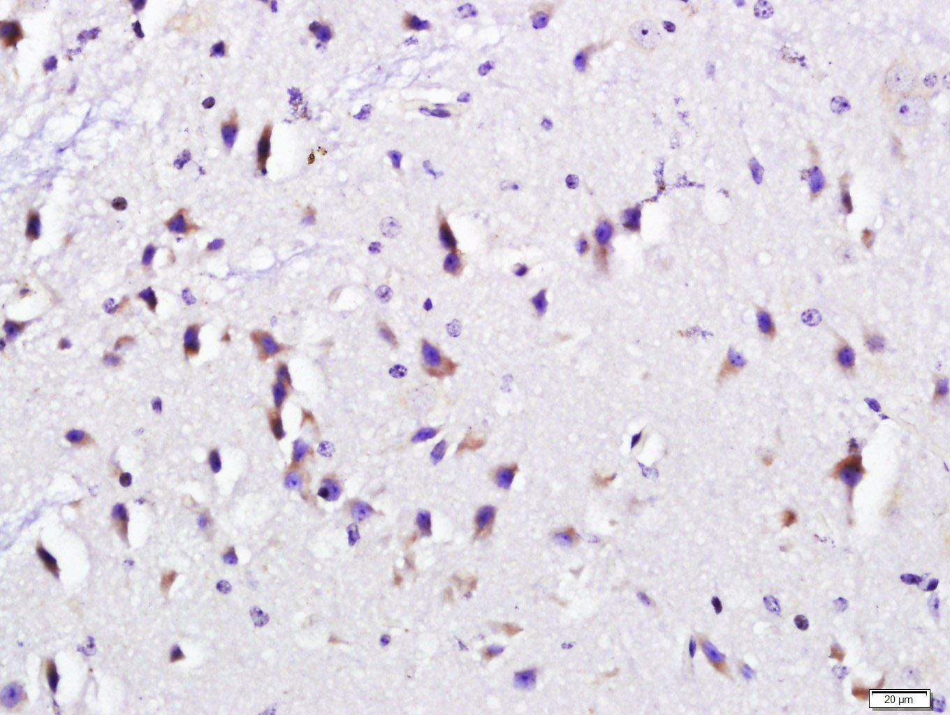
. EBAG9 was detected in a paraffin-embedded section of mouse testis tissue. Heat mediated antigen retrieval was performed in EDTA buffer (pH 8.0, epitope retrieval solution). The tissue section was blocked with 10% goat serum. The tissue section was then incubated with 1 microg/ml rabbit anti-EBAG9 Antibody (PB9553) overnight at 4°C. Biotinylated goat anti-rabbit IgG was used as secondary antibody and incubated for 30 minutes at 37°C. The tissue section was developed using Strepavidin-Biotin-Complex (SABC) (Catalog # SA1022) with DAB as the chromogen.")
. EBAG9 was detected in a paraffin-embedded section of rat testis tissue. Heat mediated antigen retrieval was performed in EDTA buffer (pH 8.0, epitope retrieval solution). The tissue section was blocked with 10% goat serum. The tissue section was then incubated with 1 microg/ml rabbit anti-EBAG9 Antibody (PB9553) overnight at 4°C. Biotinylated goat anti-rabbit IgG was used as secondary antibody and incubated for 30 minutes at 37°C. The tissue section was developed using Strepavidin-Biotin-Complex (SABC) (Catalog # SA1022) with DAB as the chromogen.")
. EBAG9 was detected in a paraffin-embedded section of human prostatic cancer tissue. Heat mediated antigen retrieval was performed in EDTA buffer (pH 8.0, epitope retrieval solution). The tissue section was blocked with 10% goat serum. The tissue section was then incubated with 1 microg/ml rabbit anti-EBAG9 Antibody (PB9553) overnight at 4°C. Biotinylated goat anti-rabbit IgG was used as secondary antibody and incubated for 30 minutes at 37°C. The tissue section was developed using Strepavidin-Biotin-Complex (SABC) (Catalog # SA1022) with DAB as the chromogen.")
Figure 1. Western blot analysis of EBAG9 using anti-EBAG9 antibody (PB9553). Electrophoresis was performed on a 5-20% SDS-PAGE gel at 70V (Stacking gel) / 90V (Resolving gel) for 2-3 hours. Lane 1: Rat Testis Tissue Lysate at 50ug, Lane 2: 22RV1 Whole Cell Lysate at 40ug, Lane 3: HELA Whole Cell Lysate at 40ug, Lane 4: MCF-7 Whole Cell Lysate at 40ug, Lane 5: JURKAT Whole Cell Lysate at 40ug. After electrophoresis, proteins were transferred to a nitrocellulose membrane at 150 mA for 50-90 minutes. Blocked the membrane with 5% non-fat milk/TBS for 1.5 hour at RT. The membrane was incubated with rabbit anti-EBAG9 antigen affinity purified polyclonal antibody (Catalog # PB9553) at 0.5 microg/mL overnight at 4°C, then washed with TBS-0.1%Tween 3 times with 5 minutes each and probed with a goat anti-rabbit IgG-HRP secondary antibody at a dilution of 1:5000 for 1.5 hour at RT. The signal is developed using an Enhanced Chemiluminescent detection (ECL) kit (Catalog # EK1002) with Tanon 5200 system. A specific band was detected for EBAG9 at approximately 34 kDa. The expected band size for EBAG9 is at 34 kDa.
Anti-EBAG9 Antibody Picoband(r)
PB9553-CARRIER-FREE
ApplicationsWestern Blot, ImmunoHistoChemistry
Product group Antibodies
ReactivityHuman, Mouse, Rat
TargetEBAG9
Overview
- SupplierBoster Bio
- Product NameAnti-EBAG9 Antibody Picoband(r)
- Delivery Days Customer9
- Application Supplier NoteTested Species: In-house tested species with positive results. By Heat: Boiling the paraffin sections in 10mM citrate buffer, pH6.0, for 20mins is required for the staining of formalin/paraffin sections. Other applications have not been tested. Optimal dilutions should be determined by end users.
- ApplicationsWestern Blot, ImmunoHistoChemistry
- CertificationResearch Use Only
- ClonalityPolyclonal
- Concentration500 ug/ml
- Gene ID9166
- Target nameEBAG9
- Target descriptionestrogen receptor binding site associated antigen 9
- Target synonymsEB9, PDAF, receptor-binding cancer antigen expressed on SiSo cells, cancer-associated surface antigen RCAS1, estrogen receptor-binding fragment-associated gene 9 protein, receptor-binding cancer antigen expressed on SiSo cells 1
- HostRabbit
- IsotypeIgG
- Protein IDO00559
- Protein NameReceptor-binding cancer antigen expressed on SiSo cells
- Scientific DescriptionBoster Bio Anti-EBAG9 Antibody Picoband® catalog # PB9553. Tested in IHC, WB applications. This antibody reacts with Human, Mouse, Rat. The brand Picoband indicates this is a premium antibody that guarantees superior quality, high affinity, and strong signals with minimal background in Western blot applications. Only our best-performing antibodies are designated as Picoband, ensuring unmatched performance.
- ReactivityHuman, Mouse, Rat
- Storage Instruction-20°C,2°C to 8°C
- UNSPSC12352203
Related products
Product group Antibodies
Anti-EBAG9 Antibody144-01935
ApplicationsWestern Blot, ImmunoHistoChemistry
ReactivityHuman, Mouse, Rat
TargetEBAG9
- SizePrice
Product group Antibodies
Anti-EBAG9 AntibodyA30526
ApplicationsWestern Blot, ImmunoHistoChemistry
ReactivityHuman, Mouse, Rat
- SizePrice
Product group Antibodies
RCAS1/EBAG9 Polyclonal AntibodyBS-10982R
ApplicationsImmunoFluorescence, ELISA, ImmunoCytoChemistry, ImmunoHistoChemistry, ImmunoHistoChemistry Frozen, ImmunoHistoChemistry Paraffin
ReactivityBovine, Chicken, Human, Mouse, Porcine, Rabbit
TargetEBAG9
- SizePrice
Product group Antibodies
EBAG9 AntibodyCSB-PA007355GA01HU
ApplicationsWestern Blot, ELISA, ImmunoHistoChemistry
ReactivityHuman, Mouse, Rat
TargetEBAG9
- SizePrice
Product group Antibodies

Goat anti-EBAG9 / RCAS1EB07292
ApplicationsWestern Blot, ELISA, ImmunoHistoChemistry
ReactivityCanine, Human, Mouse, Rat
TargetEBAG9
- SizePrice
Product group Antibodies
EBAG9 antibodyGTX134856
ApplicationsWestern Blot
ReactivityHuman
TargetEBAG9
- SizePrice
Product group Antibodies

EBAG9 / RCAS1 AntibodyLS-C335193
ApplicationsWestern Blot, ImmunoHistoChemistry
ReactivityHuman, Mouse, Rat
TargetEBAG9
- SizePrice
Product group Antibodies
Anti-EBAG9 AntibodyHPA021153
ApplicationsImmunoHistoChemistry
ReactivityHuman
TargetEBAG9
- SizePrice
Product group Antibodies
Anti-EBAG9Y058266
ApplicationsELISA, ImmunoHistoChemistry
ReactivityHuman, Mouse, Rat
- SizePrice
Product group Antibodies
Anti-EBAG9 AntibodyCAB1935
ApplicationsWestern Blot, ELISA
ReactivityHuman
TargetEBAG9
- SizePrice