


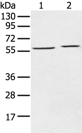
Figure 1. Western blot analysis of ECM1 using anti-ECM1 antibody (M02861-1). Electrophoresis was performed on a 5-20% SDS-PAGE gel at 70V (Stacking gel) / 90V (Resolving gel) for 2-3 hours. The sample well of each lane was loaded with 30 ug of sample under reducing conditions. Lane 1: human A431 whole cell lysates, Lane 2: human THP-1 whole cell lysates. After electrophoresis, proteins were transferred to a nitrocellulose membrane at 150 mA for 50-90 minutes. Blocked the membrane with 5% non-fat milk/TBS for 1.5 hour at RT. The membrane was incubated with rabbit anti-ECM1 antigen affinity purified monoclonal antibody (Catalog # M02861-1) at 1:500 overnight at 4°C, then washed with TBS-0.1%Tween 3 times with 5 minutes each and probed with a goat anti-rabbit IgG-HRP secondary antibody at a dilution of 1:500 for 1.5 hour at RT. The signal is developed using an Enhanced Chemiluminescent detection (ECL) kit (Catalog # EK1002) with Tanon 5200 system. A specific band was detected for ECM1 at approximately 75 kDa. The expected band size for ECM1 is at 61 kDa.
. Electrophoresis was performed on a 5-20% SDS-PAGE gel at 70V (Stacking gel) / 90V (Resolving gel) for 2-3 hours. The sample well of each lane was loaded with 30 ug of sample under reducing conditions. Lane 1: rat skin tissue lysates. After electrophoresis, proteins were transferred to a nitrocellulose membrane at 150 mA for 50-90 minutes. Blocked the membrane with 5% non-fat milk/TBS for 1.5 hour at RT. The membrane was incubated with rabbit anti-ECM1 antigen affinity purified monoclonal antibody (Catalog # M02861-1) at 1:500 overnight at 4°C, then washed with TBS-0.1%Tween 3 times with 5 minutes each and probed with a goat anti-rabbit IgG-HRP secondary antibody at a dilution of 1:500 for 1.5 hour at RT. The signal is developed using an Enhanced Chemiluminescent detection (ECL) kit (Catalog # EK1002) with Tanon 5200 system. A specific band was detected for ECM1 at approximately 75 kDa. The expected band size for ECM1 is at 61 kDa.")
. ECM1 was detected in a paraffin-embedded section of mouse heart tissue. Heat mediated antigen retrieval was performed in EDTA buffer (pH 8.0, epitope retrieval solution). The tissue section was blocked with 10% goat serum. The tissue section was then incubated with 1:50 rabbit anti-ECM1 Antibody (M02861-1) overnight at 4°C. Peroxidase Conjugated Goat Anti-rabbit IgG was used as secondary antibody and incubated for 30 minutes at 37°C. The tissue section was developed using HRP Conjugated Rabbit IgG Super Vision Assay Kit (Catalog # SV0002) with DAB as the chromogen.")
. ECM1 was detected in a paraffin-embedded section of mouse heart tissue. Heat mediated antigen retrieval was performed in EDTA buffer (pH 8.0, epitope retrieval solution). The tissue section was blocked with 10% goat serum. The tissue section was then incubated with 1:50 rabbit anti-ECM1 Antibody (M02861-1) overnight at 4°C. Peroxidase Conjugated Goat Anti-rabbit IgG was used as secondary antibody and incubated for 30 minutes at 37°C. The tissue section was developed using HRP Conjugated Rabbit IgG Super Vision Assay Kit (Catalog # SV0002) with DAB as the chromogen.")
. ECM1 was detected in a paraffin-embedded section of rat heart tissue. Heat mediated antigen retrieval was performed in EDTA buffer (pH 8.0, epitope retrieval solution). The tissue section was blocked with 10% goat serum. The tissue section was then incubated with 1:50 rabbit anti-ECM1 Antibody (M02861-1) overnight at 4°C. Peroxidase Conjugated Goat Anti-rabbit IgG was used as secondary antibody and incubated for 30 minutes at 37°C. The tissue section was developed using HRP Conjugated Rabbit IgG Super Vision Assay Kit (Catalog # SV0002) with DAB as the chromogen.")
. ECM1 was detected in a paraffin-embedded section of rat heart tissue. Heat mediated antigen retrieval was performed in EDTA buffer (pH 8.0, epitope retrieval solution). The tissue section was blocked with 10% goat serum. The tissue section was then incubated with 1:50 rabbit anti-ECM1 Antibody (M02861-1) overnight at 4°C. Peroxidase Conjugated Goat Anti-rabbit IgG was used as secondary antibody and incubated for 30 minutes at 37°C. The tissue section was developed using HRP Conjugated Rabbit IgG Super Vision Assay Kit (Catalog # SV0002) with DAB as the chromogen.")
Figure 1. Western blot analysis of ECM1 using anti-ECM1 antibody (M02861-1). Electrophoresis was performed on a 5-20% SDS-PAGE gel at 70V (Stacking gel) / 90V (Resolving gel) for 2-3 hours. The sample well of each lane was loaded with 30 ug of sample under reducing conditions. Lane 1: human A431 whole cell lysates, Lane 2: human THP-1 whole cell lysates. After electrophoresis, proteins were transferred to a nitrocellulose membrane at 150 mA for 50-90 minutes. Blocked the membrane with 5% non-fat milk/TBS for 1.5 hour at RT. The membrane was incubated with rabbit anti-ECM1 antigen affinity purified monoclonal antibody (Catalog # M02861-1) at 1:500 overnight at 4°C, then washed with TBS-0.1%Tween 3 times with 5 minutes each and probed with a goat anti-rabbit IgG-HRP secondary antibody at a dilution of 1:500 for 1.5 hour at RT. The signal is developed using an Enhanced Chemiluminescent detection (ECL) kit (Catalog # EK1002) with Tanon 5200 system. A specific band was detected for ECM1 at approximately 75 kDa. The expected band size for ECM1 is at 61 kDa.
Anti-ECM1 Rabbit Monoclonal Antibody
M02861-1
ApplicationsFlow Cytometry, ImmunoFluorescence, ImmunoPrecipitation, Western Blot, ImmunoCytoChemistry, ImmunoHistoChemistry
Product group Antibodies
ReactivityHuman, Mouse, Rat
TargetECM1
Overview
- SupplierBoster Bio
- Product NameAnti-ECM1 Rabbit Monoclonal Antibody
- Delivery Days Customer9
- ApplicationsFlow Cytometry, ImmunoFluorescence, ImmunoPrecipitation, Western Blot, ImmunoCytoChemistry, ImmunoHistoChemistry
- CertificationResearch Use Only
- ClonalityMonoclonal
- Clone ID25E30
- Gene ID1893
- Target nameECM1
- Target descriptionextracellular matrix protein 1
- Target synonymsURBWD, extracellular matrix protein 1, secretory component p85, testicular tissue protein Li 61
- HostRabbit
- IsotypeIgG
- Protein IDQ16610
- Protein NameExtracellular matrix protein 1
- Scientific DescriptionBoster Bio Anti-ECM1 Rabbit Monoclonal Antibody catalog # M02861-1. Tested in WB, IHC, ICC/IF, IP, Flow Cytometry applications. This antibody reacts with Human, Mouse, Rat.
- ReactivityHuman, Mouse, Rat
- Storage Instruction-20°C
- UNSPSC12352203
Related products
Product group Antibodies
Anti-ECM1 AntibodyA39010
ApplicationsWestern Blot, ImmunoHistoChemistry
ReactivityHuman
- SizePrice
Product group Antibodies
Anti-ECM1 Antibody144-64315
ApplicationsWestern Blot
ReactivityHuman
TargetECM1
- SizePrice
Product group Antibodies

ECM1 AntibodyLS-C831605
ApplicationsImmunoHistoChemistry
ReactivityHuman, Mouse, Rat
TargetECM1
- SizePrice
Product group Antibodies
References
ECM1 Polyclonal AntibodyBS-0776R
ApplicationsFlow Cytometry, ImmunoFluorescence, Western Blot, ELISA, ImmunoCytoChemistry, ImmunoHistoChemistry, ImmunoHistoChemistry Frozen, ImmunoHistoChemistry Paraffin
ReactivityBovine, Canine, Human, Mouse, Rat
TargetECM1
- SizePrice
Product group Antibodies
ECM1 AntibodyCSB-PA007383LA01HU
ApplicationsWestern Blot, ELISA, ImmunoHistoChemistry
ReactivityHuman
TargetECM1
- SizePrice
Product group Antibodies
ECM1 Polyclonal AntibodyCAC15356
ApplicationsWestern Blot, ELISA, ImmunoHistoChemistry
TargetECM1
- SizePrice
![IHC-P analysis of human colon carcinoma section using GTX02627 ECM1 antibody [ECM1/2889R].](https://www.genetex.com/upload/website/prouct_img/normal/GTX02627/GTX02627_20210319_IHC-P_w_23053122_685.webp)
Product group Antibodies
ECM1 antibody [ECM1/2889R]GTX02627
ApplicationsImmunoHistoChemistry, ImmunoHistoChemistry Paraffin
ReactivityHuman, Rat
TargetECM1
- SizePrice

Product group Antibodies
Anti-ECM1 AntibodyHPA027241
ApplicationsWestern Blot, ImmunoHistoChemistry
ReactivityHuman
TargetECM1
- SizePrice
Product group Antibodies
TargetECM1
- SizePrice
Product group Antibodies
Anti-ECM1 AntibodyCAB16368
ApplicationsImmunoFluorescence, Western Blot, ELISA, ImmunoCytoChemistry
ReactivityHuman
TargetECM1
- SizePrice