


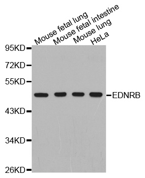
Figure 1. Western blot analysis of EDNRB using anti-EDNRB antibody (M01041-1). Electrophoresis was performed on a 5-20% SDS-PAGE gel at 70V (Stacking gel) / 90V (Resolving gel) for 2-3 hours. The sample well of each lane was loaded with 50ug of sample under reducing conditions. Lane 1: human placenta tissue lysates, Lane 2: human CACO-2 whole cell lysates, Lane 3: human SW620 whole cell lysates, Lane 4: human HepG2 whole cell lysates. After Electrophoresis, proteins were transferred to a Nitrocellulose membrane at 150mA for 50-90 minutes. Blocked the membrane with 5% Non-fat Milk/ TBS for 1.5 hour at RT. The membrane was incubated with mouse anti-EDNRB antigen affinity purified monoclonal antibody (Catalog # M01041-1) at 0.5 microg/mL overnight at 4°C, then washed with TBS-0.1%Tween 3 times with 5 minutes each and probed with a goat anti-mouse IgG-HRP secondary antibody at a dilution of 1:10000 for 1.5 hour at RT. The signal is developed using an Enhanced Chemiluminescent detection (ECL) kit (Catalog # EK1001) with Tanon 5200 system. A specific band was detected for EDNRB at approximately 45KD. The expected band size for EDNRB is at 45KD.
Figure 1. Western blot analysis of EDNRB using anti-EDNRB antibody (M01041-1). Electrophoresis was performed on a 5-20% SDS-PAGE gel at 70V (Stacking gel) / 90V (Resolving gel) for 2-3 hours. The sample well of each lane was loaded with 50ug of sample under reducing conditions. Lane 1: human placenta tissue lysates, Lane 2: human CACO-2 whole cell lysates, Lane 3: human SW620 whole cell lysates, Lane 4: human HepG2 whole cell lysates. After Electrophoresis, proteins were transferred to a Nitrocellulose membrane at 150mA for 50-90 minutes. Blocked the membrane with 5% Non-fat Milk/ TBS for 1.5 hour at RT. The membrane was incubated with mouse anti-EDNRB antigen affinity purified monoclonal antibody (Catalog # M01041-1) at 0.5 microg/mL overnight at 4°C, then washed with TBS-0.1%Tween 3 times with 5 minutes each and probed with a goat anti-mouse IgG-HRP secondary antibody at a dilution of 1:10000 for 1.5 hour at RT. The signal is developed using an Enhanced Chemiluminescent detection (ECL) kit (Catalog # EK1001) with Tanon 5200 system. A specific band was detected for EDNRB at approximately 45KD. The expected band size for EDNRB is at 45KD.
Anti-EDNRB Antibody Picoband(r) (monoclonal, 15C3)
M01041-1
ApplicationsWestern Blot
Product group Antibodies
ReactivityHuman
TargetEDNRB
Overview
- SupplierBoster Bio
- Product NameAnti-EDNRB Antibody Picoband(r) (monoclonal, 15C3)
- Delivery Days Customer9
- ApplicationsWestern Blot
- CertificationResearch Use Only
- ClonalityMonoclonal
- Clone ID15C3
- Concentration500 ug/ml
- Gene ID1910
- Target nameEDNRB
- Target descriptionendothelin receptor type B
- Target synonymsABCDS, ET-B, ET-BR, ETB, ETB1, ETBR, ETRB, HSCR, HSCR2, WS4A, endothelin receptor type B, Hirschsprung disease 2, endothelin receptor non-selective type, endothelin receptor subtype B1
- HostMouse
- IsotypeIgG2b
- Protein IDP24530
- Protein NameEndothelin receptor type B
- Scientific DescriptionBoster Bio Anti-EDNRB Antibody Picoband® (monoclonal, 15C3) catalog # M01041-1. Tested in WB applications. This antibody reacts with Human. The brand Picoband indicates this is a premium antibody that guarantees superior quality, high affinity, and strong signals with minimal background in Western blot applications. Only our best-performing antibodies are designated as Picoband, ensuring unmatched performance.
- ReactivityHuman
- Storage Instruction-20°C,2°C to 8°C
- UNSPSC12352203
References
- Gao J, Zhao L, Shahzad M, et al. Expression of endothelin-1 and its receptors in the lungs of broiler chickens exposed to high-altitude hypoxia. Avian Pathol. 2013,42(5):416-9. doi: 10.1080/03079457.2013.821568Read this paper
Datasheet
MSDS
Related products
Product group Antibodies
Anti-EDNRB AntibodyA30651
ApplicationsWestern Blot, ImmunoHistoChemistry
ReactivityHuman, Mouse
- SizePrice

Product group Antibodies
Anti-EDNRB Antibody Picoband(r)A01041-CARRIER-FREE
ApplicationsWestern Blot, ELISA
ReactivityHuman
TargetEDNRB
- SizePrice
Product group Antibodies
Anti-EDNRB Antibody144-02908
ApplicationsWestern Blot
ReactivityHuman, Mouse
TargetEDNRB
- SizePrice
Product group Antibodies
ApplicationsFlow Cytometry, Western Blot, ELISA
ReactivityBovine, Canine, Chicken, Equine, Human, Mouse, Porcine, Rabbit, Rat
TargetEDNRB
- SizePrice
Product group Antibodies
EDNRB AntibodyCSB-PA007404LA01HU
ApplicationsImmunoFluorescence, Western Blot, ELISA, ImmunoHistoChemistry
ReactivityHuman, Mouse
TargetEDNRB
- SizePrice
Product group Antibodies
EDNRB Polyclonal AntibodyCAC14720
ApplicationsImmunoFluorescence, Western Blot, ELISA, ImmunoHistoChemistry
ReactivityMouse
TargetEDNRB
- SizePrice
Product group Antibodies

EDNRB / Endothelin B Receptor AntibodyLS-C335279
ApplicationsWestern Blot
ReactivityHuman, Mouse
TargetEDNRB
- SizePrice
Product group Antibodies
Anti-EDNRB AntibodyCAB2908
ApplicationsWestern Blot, ELISA
ReactivityHuman
TargetEDNRB
- SizePrice
Product group Antibodies
TargetEDNRB
- SizePrice