


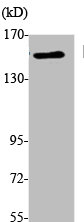
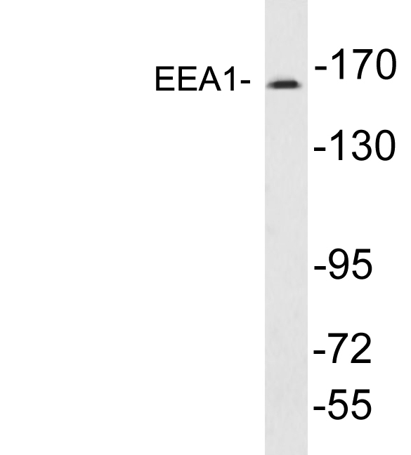
Figure 1. Western blot analysis of EEA1 using anti-EEA1 antibody (A02296-3). Electrophoresis was performed on a 5-20% SDS-PAGE gel at 70V (Stacking gel) / 90V (Resolving gel) for 2-3 hours. The sample well of each lane was loaded with 30 ug of sample under reducing conditions. Lane 1: human RT4 whole cell lysates, Lane 2: human Hacat whole cell lysates, Lane 3: human HepG2 whole cell lysates, Lane 4: human SiHa whole cell lysates, Lane 5: humna K562 whole cell lysates, Lane 6: humna MCF-7 whole cell lysates. After electrophoresis, proteins were transferred to a nitrocellulose membrane at 150 mA for 50-90 minutes. Blocked the membrane with 5% non-fat milk/TBS for 1.5 hour at RT. The membrane was incubated with rabbit anti-EEA1 antigen affinity purified polyclonal antibody (Catalog # A02296-3) at 0.5 microg/mL overnight at 4°C, then washed with TBS-0.1%Tween 3 times with 5 minutes each and probed with a goat anti-rabbit IgG-HRP secondary antibody at a dilution of 1:5000 for 1.5 hour at RT. The signal is developed using an Enhanced Chemiluminescent detection (ECL) kit (Catalog # EK1002) with Tanon 5200 system. A specific band was detected for EEA1 at approximately 162 kDa. The expected band size for EEA1 is at 162 kDa.
. Electrophoresis was performed on a 5-20% SDS-PAGE gel at 70V (Stacking gel) / 90V (Resolving gel) for 2-3 hours. The sample well of each lane was loaded with 30 ug of sample under reducing conditions. Lane 1: rat brain tissue lysates, Lane 2: rat lung tissue lysates, Lane 3: mouse brain tissue lysates, Lane 4: mouse kidney tissue lysates, Lane 5: mouse lung tissue lysates. After electrophoresis, proteins were transferred to a nitrocellulose membrane at 150 mA for 50-90 minutes. Blocked the membrane with 5% non-fat milk/TBS for 1.5 hour at RT. The membrane was incubated with rabbit anti-EEA1 antigen affinity purified polyclonal antibody (Catalog # A02296-3) at 0.5 microg/mL overnight at 4°C, then washed with TBS-0.1%Tween 3 times with 5 minutes each and probed with a goat anti-rabbit IgG-HRP secondary antibody at a dilution of 1:5000 for 1.5 hour at RT. The signal is developed using an Enhanced Chemiluminescent detection (ECL) kit (Catalog # EK1002) with Tanon 5200 system. A specific band was detected for EEA1 at approximately 162 kDa. The expected band size for EEA1 is at 162 kDa.")
. EEA1 was detected in a paraffin-embedded section of human breast cancer tissue. Heat mediated antigen retrieval was performed in EDTA buffer (pH 8.0, epitope retrieval solution). The tissue section was blocked with 10% goat serum. The tissue section was then incubated with 2 microg/ml rabbit anti-EEA1 Antibody (A02296-3) overnight at 4°C. Peroxidase Conjugated Goat Anti-rabbit IgG was used as secondary antibody and incubated for 30 minutes at 37°C. The tissue section was developed using HRP Conjugated Rabbit IgG Super Vision Assay Kit (Catalog # SV0002) with DAB as the chromogen.")
. EEA1 was detected in a paraffin-embedded section of rat brain tissue. Heat mediated antigen retrieval was performed in EDTA buffer (pH 8.0, epitope retrieval solution). The tissue section was blocked with 10% goat serum. The tissue section was then incubated with 2 microg/ml rabbit anti-EEA1 Antibody (A02296-3) overnight at 4°C. Peroxidase Conjugated Goat Anti-rabbit IgG was used as secondary antibody and incubated for 30 minutes at 37°C. The tissue section was developed using HRP Conjugated Rabbit IgG Super Vision Assay Kit (Catalog # SV0002) with DAB as the chromogen.")
. EEA1 was detected in immunocytochemical section of A549 cells. Enzyme antigen retrieval was performed using IHC enzyme antigen retrieval reagent (AR0022) for 15 mins. The cells were blocked with 10% goat serum. And then incubated with 5microg/mL rabbit anti- EEA1 Antibody (A02296-3) overnight at 4°C. DyLight®550 Conjugated Goat Anti-Rabbit IgG (BA1135) was used as secondary antibody at 1:500 dilution and incubated for 30 minutes at 37°C. The section was counterstained with DAPI. Visualize using a fluorescence microscope and filter sets appropriate for the label used.")
Figure 1. Western blot analysis of EEA1 using anti-EEA1 antibody (A02296-3). Electrophoresis was performed on a 5-20% SDS-PAGE gel at 70V (Stacking gel) / 90V (Resolving gel) for 2-3 hours. The sample well of each lane was loaded with 30 ug of sample under reducing conditions. Lane 1: human RT4 whole cell lysates, Lane 2: human Hacat whole cell lysates, Lane 3: human HepG2 whole cell lysates, Lane 4: human SiHa whole cell lysates, Lane 5: humna K562 whole cell lysates, Lane 6: humna MCF-7 whole cell lysates. After electrophoresis, proteins were transferred to a nitrocellulose membrane at 150 mA for 50-90 minutes. Blocked the membrane with 5% non-fat milk/TBS for 1.5 hour at RT. The membrane was incubated with rabbit anti-EEA1 antigen affinity purified polyclonal antibody (Catalog # A02296-3) at 0.5 microg/mL overnight at 4°C, then washed with TBS-0.1%Tween 3 times with 5 minutes each and probed with a goat anti-rabbit IgG-HRP secondary antibody at a dilution of 1:5000 for 1.5 hour at RT. The signal is developed using an Enhanced Chemiluminescent detection (ECL) kit (Catalog # EK1002) with Tanon 5200 system. A specific band was detected for EEA1 at approximately 162 kDa. The expected band size for EEA1 is at 162 kDa.
Anti-EEA1 Antibody Picoband(r)
A02296-3-CARRIER-FREE
ApplicationsImmunoFluorescence, Western Blot, ImmunoCytoChemistry, ImmunoHistoChemistry
Product group Antibodies
ReactivityHuman, Mouse, Rat
TargetEEA1
Overview
- SupplierBoster Bio
- Product NameAnti-EEA1 Antibody Picoband(r)
- Delivery Days Customer9
- ApplicationsImmunoFluorescence, Western Blot, ImmunoCytoChemistry, ImmunoHistoChemistry
- CertificationResearch Use Only
- ClonalityPolyclonal
- Concentration500 ug/ml
- Gene ID8411
- Target nameEEA1
- Target descriptionearly endosome antigen 1
- Target synonymsMST105, MSTP105, ZFYVE2, early endosome antigen 1, early endosome antigen 1, 162kD, early endosome-associated protein, endosome-associated protein p162, zinc finger FYVE domain-containing protein 2
- HostRabbit
- IsotypeIgG
- Protein IDQ15075
- Protein NameEarly endosome antigen 1
- Scientific DescriptionBoster Bio Anti-EEA1 Antibody Picoband® catalog # A02296-3. Tested in IF, IHC, ICC, WB applications. This antibody reacts with Human, Mouse, Rat. The brand Picoband indicates this is a premium antibody that guarantees superior quality, high affinity, and strong signals with minimal background in Western blot applications. Only our best-performing antibodies are designated as Picoband, ensuring unmatched performance.
- ReactivityHuman, Mouse, Rat
- Storage Instruction-20°C,2°C to 8°C
- UNSPSC12352203
Related products
Product group Antibodies
EEA1 AntibodyCSB-PA004632
ApplicationsWestern Blot, ELISA
ReactivityHuman, Mouse
TargetEEA1
- SizePrice
Product group Antibodies
Anti-EEA1 AntibodyA99427
ApplicationsWestern Blot, ELISA
ReactivityHuman, Mouse
- SizePrice
Product group Antibodies

ApplicationsImmunoFluorescence, Western Blot, ELISA, ImmunoCytoChemistry, ImmunoHistoChemistry
- SizePrice
Product group Antibodies

EEA1 AntibodyLS-C831243
ApplicationsWestern Blot, ELISA
ReactivityHuman, Mouse
TargetEEA1
- SizePrice
Product group Antibodies

Goat anti-EEA1 (aa821-835)EB11689
ApplicationsImmunoFluorescence, Western Blot, ELISA
ReactivityHuman, Mouse
TargetEEA1
- SizePrice

Product group Antibodies
Anti-EEA1 AntibodyHPA038159
ApplicationsImmunoHistoChemistry
ReactivityHuman
TargetEEA1
- SizePrice
Product group Antibodies
EEA1 Polyclonal AntibodyCAC14984
ApplicationsImmunoFluorescence, Western Blot, ELISA, ImmunoHistoChemistry
TargetEEA1
- SizePrice
![EEA1 antibody [C3], C-term detects EEA1 protein at cytosol on mouse testis by immunohistochemical analysis. Sample: Paraffin-embedded mouse testis. EEA1 antibody [C3], C-term (GTX109638) dilution: 1:500.
Antigen Retrieval: Trilogy? (EDTA based, pH 8.0) buffer, 15min](https://www.genetex.com/upload/website/prouct_img/normal/GTX109638/GTX109638_41605_IHC_M_2_w_23060500_146.webp)
Product group Antibodies
EEA1 antibody [C3], C-termGTX109638
ApplicationsImmunoFluorescence, Western Blot, ImmunoCytoChemistry, ImmunoHistoChemistry, ImmunoHistoChemistry Paraffin
ReactivityHamster, Human, Mouse, Rat
TargetEEA1
- SizePrice
Product group Antibodies
EEA1 Recombinant AntibodyBSM-61222R
ApplicationsImmunoFluorescence, Western Blot, ImmunoCytoChemistry
TargetEEA1
- SizePrice