


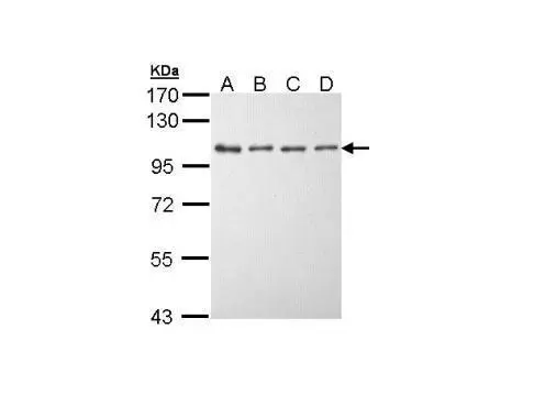
Figure 1. Western blot analysis of EEF2K using anti-EEF2K antibody (A02277-3). Electrophoresis was performed on a 5-20% SDS-PAGE gel at 70V (Stacking gel) / 90V (Resolving gel) for 2-3 hours. The sample well of each lane was loaded with 30 ug of sample under reducing conditions. Lane 1: human PC-3 whole cell lysates, Lane 2: human U-87MG whole cell lysates, Lane 3: human HepG2 whole cell lysates, Lane 4: rat C6 whole cell lysates. After electrophoresis, proteins were transferred to a nitrocellulose membrane at 150 mA for 50-90 minutes. Blocked the membrane with 5% non-fat milk/TBS for 1.5 hour at RT. The membrane was incubated with rabbit anti-EEF2K antigen affinity purified polyclonal antibody (Catalog # A02277-3) at 0.5 microg/mL overnight at 4°C, then washed with TBS-0.1%Tween 3 times with 5 minutes each and probed with a goat anti-rabbit IgG-HRP secondary antibody at a dilution of 1:5000 for 1.5 hour at RT. The signal is developed using an Enhanced Chemiluminescent detection (ECL) kit (Catalog # EK1002) with Tanon 5200 system. A specific band was detected for EEF2K at approximately 82 kDa. The expected band size for EEF2K is at 82 kDa.
. EEF2K was detected in an immunocytochemical section of HepG2 cells. Enzyme antigen retrieval was performed using IHC enzyme antigen retrieval reagent (AR0022) for 15 mins. The cells were blocked with 10% goat serum. And then incubated with 5 microg/mL rabbit anti-EEF2K Antibody (A02277-3) overnight at 4°C. DyLight®488 Conjugated Goat Anti-Rabbit IgG (BA1127) was used as secondary antibody at 1:100 dilution and incubated for 30 minutes at 37°C. The section was counterstained with DAPI. Visualize using a fluorescence microscope and filter sets appropriate for the label used.")
. Overlay histogram showing SiHa cells stained with A02277-3 (Blue line). To facilitate intracellular staining, cells were fixed with 4% paraformaldehyde and permeabilized with permeabilization buffer. The cells were blocked with 10% normal goat serum. And then incubated with rabbit anti-EEF2K Antibody (A02277-3, 1 microg/1x106 cells) for 30 min at 20°C. DyLight®488 conjugated goat anti-rabbit IgG (BA1127, 5-10 microg/1x106 cells) was used as secondary antibody for 30 minutes at 20°C. Isotype control antibody (Green line) was rabbit IgG (1 microg/1x106) used under the same conditions. Unlabelled sample without incubation with primary antibody and secondary antibody (Red line) was used as a blank control.")
Figure 1. Western blot analysis of EEF2K using anti-EEF2K antibody (A02277-3). Electrophoresis was performed on a 5-20% SDS-PAGE gel at 70V (Stacking gel) / 90V (Resolving gel) for 2-3 hours. The sample well of each lane was loaded with 30 ug of sample under reducing conditions. Lane 1: human PC-3 whole cell lysates, Lane 2: human U-87MG whole cell lysates, Lane 3: human HepG2 whole cell lysates, Lane 4: rat C6 whole cell lysates. After electrophoresis, proteins were transferred to a nitrocellulose membrane at 150 mA for 50-90 minutes. Blocked the membrane with 5% non-fat milk/TBS for 1.5 hour at RT. The membrane was incubated with rabbit anti-EEF2K antigen affinity purified polyclonal antibody (Catalog # A02277-3) at 0.5 microg/mL overnight at 4°C, then washed with TBS-0.1%Tween 3 times with 5 minutes each and probed with a goat anti-rabbit IgG-HRP secondary antibody at a dilution of 1:5000 for 1.5 hour at RT. The signal is developed using an Enhanced Chemiluminescent detection (ECL) kit (Catalog # EK1002) with Tanon 5200 system. A specific band was detected for EEF2K at approximately 82 kDa. The expected band size for EEF2K is at 82 kDa.
Anti-EEF2K Antibody Picoband(r)
A02277-3-CARRIER-FREE
ApplicationsFlow Cytometry, ImmunoFluorescence, Western Blot, ELISA, ImmunoCytoChemistry
Product group Antibodies
ReactivityHuman, Rat
TargetEEF2K
Overview
- SupplierBoster Bio
- Product NameAnti-EEF2K Antibody Picoband(r)
- Delivery Days Customer9
- ApplicationsFlow Cytometry, ImmunoFluorescence, Western Blot, ELISA, ImmunoCytoChemistry
- CertificationResearch Use Only
- ClonalityPolyclonal
- Concentration500 ug/ml
- Gene ID29904
- Target nameEEF2K
- Target descriptioneukaryotic elongation factor 2 kinase
- Target synonymsCaMKIII, HSU93850, eEF-2K, eukaryotic elongation factor 2 kinase, alternative protein EEF2K, calcium/calmodulin-dependent eukaryotic elongation factor-2 kinase, calmodulin-dependent protein kinase III, eEF-2 kinase, elongation factor-2 kinase, eukaroytic elongation factor 2 kinase
- HostRabbit
- IsotypeIgG
- Protein IDO00418
- Protein NameEukaryotic elongation factor 2 kinase
- Scientific DescriptionBoster Bio Anti-EEF2K Antibody Picoband® catalog # A02277-3. Tested in ELISA, Flow Cytometry, IF, ICC, WB applications. This antibody reacts with Human, Rat. The brand Picoband indicates this is a premium antibody that guarantees superior quality, high affinity, and strong signals with minimal background in Western blot applications. Only our best-performing antibodies are designated as Picoband, ensuring unmatched performance.
- ReactivityHuman, Rat
- Storage Instruction-20°C,2°C to 8°C
- UNSPSC12352203
Related products
Product group Antibodies
Anti-eEF2K AntibodyA97573
ApplicationsWestern Blot, ELISA
ReactivityHuman, Mouse, Rat
- SizePrice
Product group Antibodies
Anti-EEF2K Antibody144-05404
ApplicationsImmunoFluorescence, Western Blot
ReactivityHuman, Mouse
TargetEEF2K
- SizePrice
Product group Antibodies

EEF2K Antibody (Ser366)LS-C769165
ApplicationsWestern Blot, ELISA, ImmunoHistoChemistry, ImmunoHistoChemistry Paraffin
ReactivityHuman, Rat
TargetEEF2K
- SizePrice
Product group Antibodies
ApplicationsWestern Blot
ReactivityHuman, Rat
TargetEEF2K
- SizePrice
Product group Antibodies
EEF2K AntibodyCSB-PA002258
ApplicationsWestern Blot, ELISA, ImmunoHistoChemistry
ReactivityHuman, Rat
TargetEEF2K
- SizePrice
Product group Antibodies
Eef2K Polyclonal AntibodyCAC08385
ApplicationsImmunoFluorescence, ELISA, ImmunoHistoChemistry
TargetEEF2K
- SizePrice

Product group Antibodies
Anti-EEF2K AntibodyHPA056061
ApplicationsImmunoCytoChemistry
ReactivityHuman
TargetEEF2K
- SizePrice
Product group Antibodies
eEF2K antibody [N1N3]GTX111496
ApplicationsWestern Blot
ReactivityHuman
TargetEEF2K
- SizePrice
Product group Antibodies
ApplicationsImmunoFluorescence, Western Blot, ELISA, ImmunoCytoChemistry
ReactivityHuman
TargetEEF2K
- SizePrice