

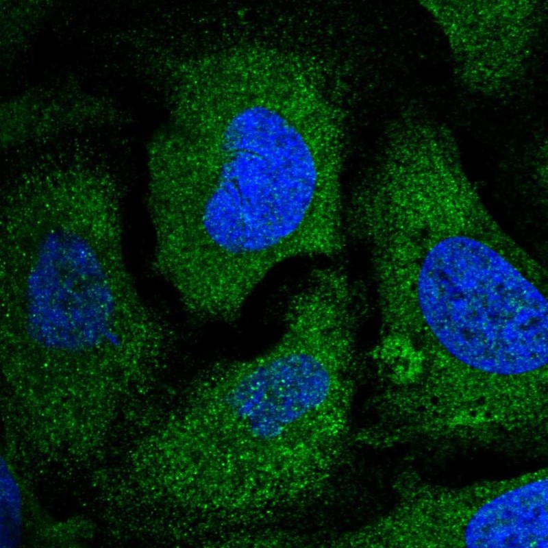
Immunofluorescent staining of human cell line U-2 OS shows localization to endoplasmic reticulum.
Immunofluorescent staining of human cell line U-2 OS shows localization to endoplasmic reticulum.
Anti-EIF2S2 Antibody
HPA041262
ApplicationsImmunoCytoChemistry
Product group Antibodies
ReactivityHuman
TargetEIF2S2
Overview
- SupplierAtlas Antibodies
- Product NameAnti-EIF2S2 Antibody
- Delivery Days Customer4
- ApplicationsImmunoCytoChemistry
- CertificationResearch Use Only
- ClonalityPolyclonal
- ConjugateUnconjugated
- Gene ID8894
- Target nameEIF2S2
- Target descriptioneukaryotic translation initiation factor 2 subunit beta
- Target synonymsEIF2, EIF2B, EIF2beta, PPP1R67, eIF-2-beta, eukaryotic translation initiation factor 2 subunit 2, eIF2-beta, eukaryotic translation initiation factor 2, subunit 2 beta, 38kDa, protein phosphatase 1, regulatory subunit 67
- HostRabbit
- IsotypeIgG
- Protein IDP20042
- Protein NameEukaryotic translation initiation factor 2 subunit 2
- Scientific DescriptionRecombinant Protein Epitope Signature Tag (PrEST) antigen sequence
- ReactivityHuman
- Storage Instruction-20°C,2°C to 8°C
- UNSPSC41116161
Datasheet
MSDS
Related products
Product group Antibodies
EIF2S2 AntibodyCSB-PA010574
ApplicationsWestern Blot, ELISA, ImmunoHistoChemistry
ReactivityHuman, Mouse
TargetEIF2S2
- SizePrice
Product group Antibodies
Anti-EIF2S2 AntibodyA100577
ApplicationsWestern Blot, ELISA
ReactivityHuman
- SizePrice
Product group Antibodies

EIF2S2 AntibodyLS-C760952
ApplicationsWestern Blot
ReactivityHuman, Mouse, Rat
TargetEIF2S2
- SizePrice
Product group Antibodies
Anti-EIF2S2 AntibodyHPA041332
ApplicationsImmunoCytoChemistry, ImmunoHistoChemistry
ReactivityHuman
TargetEIF2S2
- SizePrice

Product group Antibodies
Anti-EIF2S2 Antibody Picoband(r)PA2157-CARRIER-FREE
ApplicationsWestern Blot
ReactivityHamster, Human
TargetEIF2S2
- SizePrice
Product group Antibodies
EIF2 beta antibodyGTX106484
ApplicationsImmunoFluorescence, ImmunoPrecipitation, Western Blot, ImmunoCytoChemistry, ImmunoHistoChemistry, ImmunoHistoChemistry Paraffin
ReactivityHuman, Mouse, Rat
TargetEIF2S2
- SizePrice