


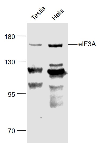
Figure 1. Western blot analysis of EIF3A using anti-EIF3A antibody (A02339-3). Electrophoresis was performed on a 5-20% SDS-PAGE gel at 70V (Stacking gel) / 90V (Resolving gel) for 2-3 hours. The sample well of each lane was loaded with 30 ug of sample under reducing conditions. Lane 1: human PC-3 whole cell lysates, Lane 2: human 293T whole cell lysates, Lane 3: human MOLT-4 whole cell lysates, Lane 4: rat C6 whole cell lysates, Lane 5: rat L6 whole cell lysates, Lane 6: mouse RAW264.7 whole cell lysates, Lane 7: mouse ANA-1 whole cell lysates. After electrophoresis, proteins were transferred to a nitrocellulose membrane at 150 mA for 50-90 minutes. Blocked the membrane with 5% non-fat milk/TBS for 1.5 hour at RT. The membrane was incubated with rabbit anti-EIF3A antigen affinity purified polyclonal antibody (Catalog # A02339-3) at 0.5 microg/mL overnight at 4°C, then washed with TBS-0.1%Tween 3 times with 5 minutes each and probed with a goat anti-rabbit IgG-HRP secondary antibody at a dilution of 1:5000 for 1.5 hour at RT. The signal is developed using an Enhanced Chemiluminescent detection (ECL) kit (Catalog # EK1002) with Tanon 5200 system. A specific band was detected for EIF3A at approximately 180 kDa. The expected band size for EIF3A is at 167 kDa.
. EIF3A was detected in a paraffin-embedded section of human colorectal adenocarcinoma tissue. Heat mediated antigen retrieval was performed in EDTA buffer (pH 8.0, epitope retrieval solution). The tissue section was blocked with 10% goat serum. The tissue section was then incubated with 2 microg/ml rabbit anti-EIF3A Antibody (A02339-3) overnight at 4°C. Peroxidase Conjugated Goat Anti-rabbit IgG was used as secondary antibody and incubated for 30 minutes at 37°C. The tissue section was developed using HRP Conjugated Rabbit IgG Super Vision Assay Kit (Catalog # SV0002) with DAB as the chromogen.")
. EIF3A was detected in a paraffin-embedded section of human esophageal squamous carcinoma tissue. Heat mediated antigen retrieval was performed in EDTA buffer (pH 8.0, epitope retrieval solution). The tissue section was blocked with 10% goat serum. The tissue section was then incubated with 2 microg/ml rabbit anti-EIF3A Antibody (A02339-3) overnight at 4°C. Peroxidase Conjugated Goat Anti-rabbit IgG was used as secondary antibody and incubated for 30 minutes at 37°C. The tissue section was developed using HRP Conjugated Rabbit IgG Super Vision Assay Kit (Catalog # SV0002) with DAB as the chromogen.")
. EIF3A was detected in a paraffin-embedded section of mouse brain tissue. Heat mediated antigen retrieval was performed in EDTA buffer (pH 8.0, epitope retrieval solution). The tissue section was blocked with 10% goat serum. The tissue section was then incubated with 2 microg/ml rabbit anti-EIF3A Antibody (A02339-3) overnight at 4°C. Peroxidase Conjugated Goat Anti-rabbit IgG was used as secondary antibody and incubated for 30 minutes at 37°C. The tissue section was developed using HRP Conjugated Rabbit IgG Super Vision Assay Kit (Catalog # SV0002) with DAB as the chromogen.")
. EIF3A was detected in a paraffin-embedded section of rat brain tissue. Heat mediated antigen retrieval was performed in EDTA buffer (pH 8.0, epitope retrieval solution). The tissue section was blocked with 10% goat serum. The tissue section was then incubated with 2 microg/ml rabbit anti-EIF3A Antibody (A02339-3) overnight at 4°C. Peroxidase Conjugated Goat Anti-rabbit IgG was used as secondary antibody and incubated for 30 minutes at 37°C. The tissue section was developed using HRP Conjugated Rabbit IgG Super Vision Assay Kit (Catalog # SV0002) with DAB as the chromogen.")
. Overlay histogram showing U87 cells stained with A02339-3 (Blue line). To facilitate intracellular staining, cells were fixed with 4% paraformaldehyde and permeabilized with permeabilization buffer. The cells were blocked with 10% normal goat serum. And then incubated with rabbit anti-EIF3A Antibody (A02339-3, 1 microg/1x106 cells) for 30 min at 20°C. DyLight®488 conjugated goat anti-rabbit IgG (BA1127, 5-10 microg/1x106 cells) was used as secondary antibody for 30 minutes at 20°C. Isotype control antibody (Green line) was rabbit IgG (1 microg/1x106) used under the same conditions. Unlabelled sample without incubation with primary antibody and secondary antibody (Red line) was used as a blank control.")
. EIF3A was detected in a paraffin-embedded section of human intestinal cancer tissue. Heat mediated antigen retrieval was performed in EDTA buffer (pH 8.0, epitope retrieval solution). The tissue section was blocked with 10% goat serum. The tissue section was then incubated with 5 microg/mL rabbit anti-EIF3A Antibody (A02339-3) overnight at 4°C. DyLight®550 Conjugated Goat Anti-Rabbit IgG (BA1135) was used as secondary antibody at 1:500 dilution and incubated for 30 minutes at 37°C. The section was counterstained with DAPI. Visualize using a fluorescence microscope and filter sets appropriate for the label used.")
Figure 1. Western blot analysis of EIF3A using anti-EIF3A antibody (A02339-3). Electrophoresis was performed on a 5-20% SDS-PAGE gel at 70V (Stacking gel) / 90V (Resolving gel) for 2-3 hours. The sample well of each lane was loaded with 30 ug of sample under reducing conditions. Lane 1: human PC-3 whole cell lysates, Lane 2: human 293T whole cell lysates, Lane 3: human MOLT-4 whole cell lysates, Lane 4: rat C6 whole cell lysates, Lane 5: rat L6 whole cell lysates, Lane 6: mouse RAW264.7 whole cell lysates, Lane 7: mouse ANA-1 whole cell lysates. After electrophoresis, proteins were transferred to a nitrocellulose membrane at 150 mA for 50-90 minutes. Blocked the membrane with 5% non-fat milk/TBS for 1.5 hour at RT. The membrane was incubated with rabbit anti-EIF3A antigen affinity purified polyclonal antibody (Catalog # A02339-3) at 0.5 microg/mL overnight at 4°C, then washed with TBS-0.1%Tween 3 times with 5 minutes each and probed with a goat anti-rabbit IgG-HRP secondary antibody at a dilution of 1:5000 for 1.5 hour at RT. The signal is developed using an Enhanced Chemiluminescent detection (ECL) kit (Catalog # EK1002) with Tanon 5200 system. A specific band was detected for EIF3A at approximately 180 kDa. The expected band size for EIF3A is at 167 kDa.
Anti-EIF3A Antibody Picoband(r)
A02339-3-CARRIER-FREE
ApplicationsFlow Cytometry, ImmunoFluorescence, Western Blot, ELISA, ImmunoHistoChemistry
Product group Antibodies
ReactivityHuman, Mouse, Rat
TargetEIF3A
Overview
- SupplierBoster Bio
- Product NameAnti-EIF3A Antibody Picoband(r)
- Delivery Days Customer9
- ApplicationsFlow Cytometry, ImmunoFluorescence, Western Blot, ELISA, ImmunoHistoChemistry
- CertificationResearch Use Only
- ClonalityPolyclonal
- Concentration500 ug/ml
- Gene ID8661
- Target nameEIF3A
- Target descriptioneukaryotic translation initiation factor 3 subunit A
- Target synonymsEIF3, EIF3S10, P167, TIF32, eIF3-p170, eIF3-theta, p180, p185, eukaryotic translation initiation factor 3 subunit A, EIF3, p180 subunit, centrosomin homolog, cytoplasmic protein p167, eIF-3-theta, eIF3 p167, eIF3 p180, eIF3 p185, eukaryotic translation initiation factor 3 subunit 10, eukaryotic translation initiation factor 3, subunit 10 (theta, 150/170kD), eukaryotic translation initiation factor 3, subunit 10 (theta, 170kD), eukaryotic translation initiation factor 3, subunit 10 theta, 150/170kDa, eukaryotic translation initiation factor 3, subunit 10, 170kD
- HostRabbit
- IsotypeIgG
- Protein IDQ14152
- Protein NameEukaryotic translation initiation factor 3 subunit A
- Scientific DescriptionBoster Bio Anti-EIF3A Antibody Picoband® catalog # A02339-3. Tested in ELISA, Flow Cytometry, IF, IHC, WB applications. This antibody reacts with Human, Mouse, Rat. The brand Picoband indicates this is a premium antibody that guarantees superior quality, high affinity, and strong signals with minimal background in Western blot applications. Only our best-performing antibodies are designated as Picoband, ensuring unmatched performance.
- ReactivityHuman, Mouse, Rat
- Storage Instruction-20°C,2°C to 8°C
- UNSPSC12352203
Related products
Product group Antibodies
Anti-EIF3A Antibody144-00573
ApplicationsImmunoFluorescence, Western Blot, ImmunoHistoChemistry
ReactivityHuman, Mouse, Rat
TargetEIF3A
- SizePrice
Product group Antibodies
eIF3A Polyclonal AntibodyBS-3840R
ApplicationsImmunoFluorescence, Western Blot, ELISA, ImmunoCytoChemistry, ImmunoHistoChemistry, ImmunoHistoChemistry Frozen, ImmunoHistoChemistry Paraffin
ReactivityBovine, Canine, Equine, Human, Mouse, Porcine, Rabbit, Rat
TargetEIF3A
- SizePrice
Product group Antibodies
EIF3A AntibodyCSB-PA623903LA01HU
ApplicationsImmunoFluorescence, Western Blot, ELISA
ReactivityHuman
TargetEIF3A
- SizePrice
Product group Antibodies
EIF3A Polyclonal AntibodyCAC14900
ApplicationsImmunoFluorescence, Western Blot, ELISA
TargetEIF3A
- SizePrice
Product group Antibodies

EIF3A AntibodyLS-C408226
ApplicationsImmunoFluorescence, Western Blot, ImmunoHistoChemistry
ReactivityHuman, Mouse, Rat
TargetEIF3A
- SizePrice
Product group Antibodies
EIF3A antibody [N1N2], N-termGTX118895
ApplicationsImmunoFluorescence, Western Blot, ImmunoCytoChemistry, ImmunoHistoChemistry, ImmunoHistoChemistry Paraffin
ReactivityHuman
TargetEIF3A
- SizePrice

Product group Antibodies
Anti-EIF3A AntibodyHPA038315
ApplicationsWestern Blot, ImmunoHistoChemistry
ReactivityHuman, Mouse, Rat
TargetEIF3A
- SizePrice
Product group Antibodies
Anti-EIF3A AntibodyCAB0573
ApplicationsImmunoFluorescence, Western Blot, ELISA, ImmunoCytoChemistry
ReactivityHuman
TargetEIF3A
- SizePrice