

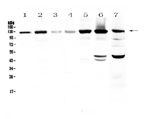
Figure 1. Western blot analysis of Eph receptor A2 using anti-Eph receptor A2 antibody (A00578). Electrophoresis was performed on a 5-20% SDS-PAGE gel at 70V (Stacking gel) / 90V (Resolving gel) for 2-3 hours. The sample well of each lane was loaded with 50ug of sample under reducing conditions. Lane 1: human Hela cell lysate, Lane 2: human U-87MG cell lysate, Lane 3: human SHG-44 cell lysate, Lane 4: human COLO-320 cell lysate, Lane 5: human SK-OV-3 cell lysate, Lane 6: human A549 cell lysate, Lane 7: mouse HEPA1-6 cell lysate. After Electrophoresis, proteins were transferred to a Nitrocellulose membrane at 150mA for 50-90 minutes. Blocked the membrane with 5% Non-fat Milk/ TBS for 1.5 hour at RT. The membrane was incubated with rabbit anti-Eph receptor A2 antigen affinity purified polyclonal antibody (Catalog # A00578) at 0.5 microg/mL overnight at 4°C, then washed with TBS-0.1%Tween 3 times with 5 minutes each and probed with a goat anti-rabbit IgG-HRP secondary antibody at a dilution of 1:10000 for 1.5 hour at RT. The signal is developed using an Enhanced Chemiluminescent detection (ECL) kit (Catalog # EK1002) with Tanon 5200 system. A specific band was detected for Eph receptor A2 at approximately 125KD. The expected band size for Eph receptor A2 is at 108KD.
. Overlay histogram showing A549 cells stained with A00578 (Blue line). To facilitate intracellular staining, cells were fixed with 4% paraformaldehyde and permeabilized with permeabilization buffer. The cells were blocked with 10% normal goat serum. And then incubated with rabbit anti-Eph receptor A2 Antibody (A00578,1microg/1x106 cells) for 30 min at 20°C. DyLight®488 conjugated goat anti-rabbit IgG (BA1127, 5-10microg/1x106 cells) was used as secondary antibody for 30 minutes at 20°C. Isotype control antibody (Green line) was rabbit IgG (1microg/1x106) used under the same conditions. Unlabelled sample without incubation with primary antibody and secondary antibody (Red line) was used as a blank control.")
. Overlay histogram showing U20S cells stained with A00578 (Blue line). To facilitate intracellular staining, cells were fixed with 4% paraformaldehyde and permeabilized with permeabilization buffer. The cells were blocked with 10% normal goat serum. And then incubated with rabbit anti-Eph receptor A2 Antibody (A00578,1microg/1x106 cells) for 30 min at 20°C. DyLight®488 conjugated goat anti-rabbit IgG (BA1127, 5-10microg/1x106 cells) was used as secondary antibody for 30 minutes at 20°C. Isotype control antibody (Green line) was rabbit IgG (1microg/1x106) used under the same conditions. Unlabelled sample without incubation with primary antibody and secondary antibody (Red line) was used as a blank control.")
. Eph receptor A2 was detected in immunocytochemical section of PC-3 cells. Enzyme antigen retrieval was performed using IHC enzyme antigen retrieval reagent (AR0022) for 15 mins. The cells were blocked with 10% goat serum. And then incubated with 5microg/mL rabbit anti-Eph receptor A2 Antibody (A00578) overnight at 4°C. DyLight®488 Conjugated Goat Anti-Rabbit IgG (BA1127) was used as secondary antibody at 1:100 dilution and incubated for 30 minutes at 37°C. The section was counterstained with DAPI. Visualize using a fluorescence microscope and filter sets appropriate for the label used.")
Figure 1. Western blot analysis of Eph receptor A2 using anti-Eph receptor A2 antibody (A00578). Electrophoresis was performed on a 5-20% SDS-PAGE gel at 70V (Stacking gel) / 90V (Resolving gel) for 2-3 hours. The sample well of each lane was loaded with 50ug of sample under reducing conditions. Lane 1: human Hela cell lysate, Lane 2: human U-87MG cell lysate, Lane 3: human SHG-44 cell lysate, Lane 4: human COLO-320 cell lysate, Lane 5: human SK-OV-3 cell lysate, Lane 6: human A549 cell lysate, Lane 7: mouse HEPA1-6 cell lysate. After Electrophoresis, proteins were transferred to a Nitrocellulose membrane at 150mA for 50-90 minutes. Blocked the membrane with 5% Non-fat Milk/ TBS for 1.5 hour at RT. The membrane was incubated with rabbit anti-Eph receptor A2 antigen affinity purified polyclonal antibody (Catalog # A00578) at 0.5 microg/mL overnight at 4°C, then washed with TBS-0.1%Tween 3 times with 5 minutes each and probed with a goat anti-rabbit IgG-HRP secondary antibody at a dilution of 1:10000 for 1.5 hour at RT. The signal is developed using an Enhanced Chemiluminescent detection (ECL) kit (Catalog # EK1002) with Tanon 5200 system. A specific band was detected for Eph receptor A2 at approximately 125KD. The expected band size for Eph receptor A2 is at 108KD.
Anti-Eph receptor A2/EPHA2 Antibody Picoband(r)
A00578-CARRIER-FREE
ApplicationsFlow Cytometry, ImmunoFluorescence, Western Blot, ELISA, ImmunoCytoChemistry
Product group Antibodies
ReactivityHuman, Mouse, Rat
TargetEPHA2
Overview
- SupplierBoster Bio
- Product NameAnti-Eph receptor A2/EPHA2 Antibody Picoband(r)
- Delivery Days Customer9
- ApplicationsFlow Cytometry, ImmunoFluorescence, Western Blot, ELISA, ImmunoCytoChemistry
- CertificationResearch Use Only
- ClonalityPolyclonal
- Concentration500 ug/ml
- Gene ID1969
- Target nameEPHA2
- Target descriptionEPH receptor A2
- Target synonymsARCC2, CTPA, CTPP1, CTRCT6, ECK, ephrin type-A receptor 2, epithelial cell receptor protein tyrosine kinase, tyrosine-protein kinase receptor ECK
- HostRabbit
- IsotypeIgG
- Protein IDP29317
- Protein NameEphrin type-A receptor 2
- Scientific DescriptionBoster Bio Anti-Eph receptor A2/EPHA2 Antibody Picoband® catalog # A00578. Tested in ELISA, Flow Cytometry, IF, ICC, WB applications. This antibody reacts with Human, Mouse, Rat. The brand Picoband indicates this is a premium antibody that guarantees superior quality, high affinity, and strong signals with minimal background in Western blot applications. Only our best-performing antibodies are designated as Picoband, ensuring unmatched performance.
- ReactivityHuman, Mouse, Rat
- Storage Instruction-20°C,2°C to 8°C
- UNSPSC12352203
Related products
Product group Antibodies
Anti-EPHA2 AntibodyA99110
ApplicationsWestern Blot, ELISA
ReactivityHuman, Mouse
- SizePrice
Product group Antibodies

Anti-Human Ephrin Type A receptor 2 [1C1]Ab00430-1.1
ApplicationsFlow Cytometry, ImmunoFluorescence, Western Blot, ELISA, Other Application
ReactivityHuman, Mouse
TargetEPHA2
- SizePrice
Product group Antibodies
Anti-EPHA2 Antibody144-07183
ApplicationsWestern Blot, ImmunoHistoChemistry
ReactivityHuman, Mouse, Rat
TargetEPHA2
- SizePrice
Product group Antibodies
References
EphA2 Polyclonal AntibodyBS-0485R
ApplicationsImmunoFluorescence, Western Blot, ELISA, ImmunoHistoChemistry, ImmunoHistoChemistry Frozen, ImmunoHistoChemistry Paraffin
ReactivityBovine, Canine, Human, Monkey, Mouse, Rat
TargetEPHA2
- SizePrice
Product group Antibodies
EPHA2/EPHA3/EPHA4 AntibodyCSB-PA002362
ApplicationsImmunoFluorescence, Western Blot, ELISA
ReactivityHuman, Rat
TargetEPHA2
- SizePrice
Product group Antibodies

EPHA2 / EPH Receptor A2 AntibodyLS-C349049
ApplicationsWestern Blot, ImmunoHistoChemistry
ReactivityHuman, Mouse, Rat
TargetEPHA2
- SizePrice
Product group Antibodies
References
EphA2 antibodyGTX32587
ApplicationsWestern Blot, ImmunoHistoChemistry, ImmunoHistoChemistry Paraffin
ReactivityHuman, Mouse, Rat
TargetEPHA2
- SizePrice
Product group Antibodies
Anti-EPHA2 AntibodyCAB7183
ApplicationsWestern Blot, ELISA
ReactivityHuman
TargetEPHA2
- SizePrice