

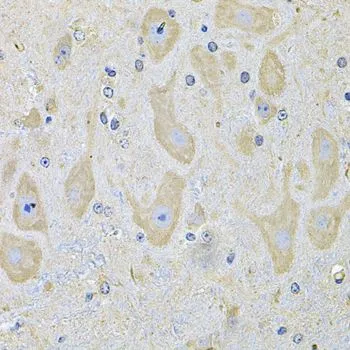
IHC-P analysis of mouse brain tissue using GTX32587 EphA2 antibody. Dilution : 1:100


IHC-P analysis of mouse brain tissue using GTX32587 EphA2 antibody. Dilution : 1:100
EphA2 antibody
GTX32587
ApplicationsWestern Blot, ImmunoHistoChemistry, ImmunoHistoChemistry Paraffin
Product group Antibodies
ReactivityHuman, Mouse, Rat
TargetEPHA2
Overview
- SupplierGeneTex
- Product NameEphA2 antibody
- Delivery Days Customer9
- Application Supplier NoteWB: 1:500 - 1:2000. IHC-P: 1:50 - 1:200. *Optimal dilutions/concentrations should be determined by the researcher.Not tested in other applications.
- ApplicationsWestern Blot, ImmunoHistoChemistry, ImmunoHistoChemistry Paraffin
- CertificationResearch Use Only
- ClonalityPolyclonal
- ConjugateUnconjugated
- Gene ID1969
- Target nameEPHA2
- Target descriptionEPH receptor A2
- Target synonymsARCC2, CTPA, CTPP1, CTRCT6, ECK, ephrin type-A receptor 2, epithelial cell receptor protein tyrosine kinase, tyrosine-protein kinase receptor ECK
- HostRabbit
- IsotypeIgG
- Protein IDP29317
- Protein NameEphrin type-A receptor 2
- Scientific DescriptionThis gene belongs to the ephrin receptor subfamily of the protein-tyrosine kinase family. EPH and EPH-related receptors have been implicated in mediating developmental events, particularly in the nervous system. Receptors in the EPH subfamily typically have a single kinase domain and an extracellular region containing a Cys-rich domain and 2 fibronectin type III repeats. The ephrin receptors are divided into 2 groups based on the similarity of their extracellular domain sequences and their affinities for binding ephrin-A and ephrin-B ligands. This gene encodes a protein that binds ephrin-A ligands. Mutations in this gene are the cause of certain genetically-related cataract disorders.[provided by RefSeq, May 2010]
- ReactivityHuman, Mouse, Rat
- Storage Instruction-20°C or -80°C,2°C to 8°C
- UNSPSC41116161
References
- Tissue Engineering of Axially Vascularized Soft-Tissue Flaps with a Poly-(epsilon-Caprolactone) Nanofiber-Hydrogel Composite. Henn D et al., 2020 Jul 1, Adv Wound Care (New Rochelle)Read this paper
Datasheet
Related products
Product group Antibodies
Anti-EPHA2 AntibodyA99110
ApplicationsWestern Blot, ELISA
ReactivityHuman, Mouse
- SizePrice
Product group Antibodies

Anti-Human Ephrin Type A receptor 2 [1C1]Ab00430-1.1
ApplicationsFlow Cytometry, ImmunoFluorescence, Western Blot, ELISA, Other Application
ReactivityHuman, Mouse
TargetEPHA2
- SizePrice
Product group Antibodies
Anti-EPHA2 Antibody144-07183
ApplicationsWestern Blot, ImmunoHistoChemistry
ReactivityHuman, Mouse, Rat
TargetEPHA2
- SizePrice
Product group Antibodies
Anti-Eph receptor A2/EPHA2 Antibody Picoband(r)A00578-CARRIER-FREE
ApplicationsFlow Cytometry, ImmunoFluorescence, Western Blot, ELISA, ImmunoCytoChemistry
ReactivityHuman, Mouse, Rat
TargetEPHA2
- SizePrice
Product group Antibodies
References
EphA2 Polyclonal AntibodyBS-0485R
ApplicationsImmunoFluorescence, Western Blot, ELISA, ImmunoHistoChemistry, ImmunoHistoChemistry Frozen, ImmunoHistoChemistry Paraffin
ReactivityBovine, Canine, Human, Monkey, Mouse, Rat
TargetEPHA2
- SizePrice
Product group Antibodies
EPHA2/EPHA3/EPHA4 AntibodyCSB-PA002362
ApplicationsImmunoFluorescence, Western Blot, ELISA
ReactivityHuman, Rat
TargetEPHA2
- SizePrice
Product group Antibodies

EPHA2 / EPH Receptor A2 AntibodyLS-C349049
ApplicationsWestern Blot, ImmunoHistoChemistry
ReactivityHuman, Mouse, Rat
TargetEPHA2
- SizePrice
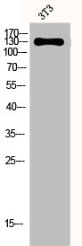
![WB analysis of NIH3T3 cell lysate using GTX83159 EphA2 antibody [1B3C7].](https://www.genetex.com/upload/website/prouct_img/normal/GTX83159/GTX83159_20170912_WB_w_23061322_688.webp)
Product group Antibodies
EphA2 antibody [1B3C7]GTX83159
ApplicationsWestern Blot, ELISA, ImmunoHistoChemistry, ImmunoHistoChemistry Paraffin
ReactivityHuman, Mouse
TargetEPHA2
- SizePrice
Product group Antibodies
EphA2 antibodyGTX56226
ApplicationsWestern Blot
ReactivityHuman
TargetEPHA2
- SizePrice