


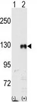
Figure 1. Western blot analysis of Eph receptor B1 using anti-Eph receptor B1 antibody (PB9584). Electrophoresis was performed on a 5-20% SDS-PAGE gel at 70V (Stacking gel) / 90V (Resolving gel) for 2-3 hours. The sample well of each lane was loaded with 30 ug of sample under reducing conditions. Lane 1: human U251 whole cell lysates, Lane 2: human Hel whole cell lysates, Lane 3: rat testis tissue lysates, Lane 4: rat brain tissue lysates, Lane 5: mouse testis tissue lysates, Lane 6: mouse brain tissue lysates. After electrophoresis, proteins were transferred to a nitrocellulose membrane at 150 mA for 50-90 minutes. Blocked the membrane with 5% non-fat milk/TBS for 1.5 hour at RT. The membrane was incubated with rabbit anti-Eph receptor B1 antigen affinity purified polyclonal antibody (Catalog # PB9584) at 0.5 microg/mL overnight at 4°C, then washed with TBS-0.1%Tween 3 times with 5 minutes each and probed with a goat anti-rabbit IgG-HRP secondary antibody at a dilution of 1:5000 for 1.5 hour at RT. The signal is developed using an Enhanced Chemiluminescent detection (ECL) kit (Catalog # EK1002) with Tanon 5200 system. A specific band was detected for Eph receptor B1 at approximately 120 kDa. The expected band size for Eph receptor B1 is at 110 kDa.
. Eph receptor B1 was detected in paraffin-embedded section of Human Glioma Tissue. Heat mediated antigen retrieval was performed in citrate buffer (pH6, epitope retrieval solution) for 20 mins. The tissue section was blocked with 10% goat serum. The tissue section was then incubated with 2microg/ml rabbit anti-Eph receptor B1 Antibody (PB9584) overnight at 4°C. Biotinylated goat anti-rabbit IgG was used as secondary antibody and incubated for 30 minutes at 37°C. The tissue section was developed using Strepavidin-Biotin-Complex (SABC)(Catalog # SA1022) with DAB as the chromogen.")
. Overlay histogram showing U20S cells stained with PB9584 (Blue line). To facilitate intracellular staining, cells were fixed with 4% paraformaldehyde and permeabilized with permeabilization buffer. The cells were blocked with 10% normal goat serum. And then incubated with rabbit anti-Eph receptor B1 Antibody (PB9584,1microg/1x106 cells) for 30 min at 20°C. DyLight®488 conjugated goat anti-rabbit IgG (BA1127, 5-10microg/1x106 cells) was used as secondary antibody for 30 minutes at 20°C. Isotype control antibody (Green line) was rabbit IgG (1microg/1x106) used under the same conditions. Unlabelled sample without incubation with primary antibody and secondary antibody (Red line) was used as a blank control.")
Figure 1. Western blot analysis of Eph receptor B1 using anti-Eph receptor B1 antibody (PB9584). Electrophoresis was performed on a 5-20% SDS-PAGE gel at 70V (Stacking gel) / 90V (Resolving gel) for 2-3 hours. The sample well of each lane was loaded with 30 ug of sample under reducing conditions. Lane 1: human U251 whole cell lysates, Lane 2: human Hel whole cell lysates, Lane 3: rat testis tissue lysates, Lane 4: rat brain tissue lysates, Lane 5: mouse testis tissue lysates, Lane 6: mouse brain tissue lysates. After electrophoresis, proteins were transferred to a nitrocellulose membrane at 150 mA for 50-90 minutes. Blocked the membrane with 5% non-fat milk/TBS for 1.5 hour at RT. The membrane was incubated with rabbit anti-Eph receptor B1 antigen affinity purified polyclonal antibody (Catalog # PB9584) at 0.5 microg/mL overnight at 4°C, then washed with TBS-0.1%Tween 3 times with 5 minutes each and probed with a goat anti-rabbit IgG-HRP secondary antibody at a dilution of 1:5000 for 1.5 hour at RT. The signal is developed using an Enhanced Chemiluminescent detection (ECL) kit (Catalog # EK1002) with Tanon 5200 system. A specific band was detected for Eph receptor B1 at approximately 120 kDa. The expected band size for Eph receptor B1 is at 110 kDa.
Anti-Eph receptor B1/EPHB1 Antibody Picoband(r)
PB9584-CARRIER-FREE
ApplicationsFlow Cytometry, Western Blot, ImmunoHistoChemistry
Product group Antibodies
ReactivityHamster, Human, Mouse, Rat
TargetEPHB1
Overview
- SupplierBoster Bio
- Product NameAnti-Eph receptor B1/EPHB1 Antibody Picoband(r)
- Delivery Days Customer9
- Application Supplier NoteTested Species: In-house tested species with positive results. By Heat: Boiling the paraffin sections in 10mM citrate buffer, pH6.0, for 20mins is required for the staining of formalin/paraffin sections. Other applications have not been tested. Optimal dilutions should be determined by end users.
- ApplicationsFlow Cytometry, Western Blot, ImmunoHistoChemistry
- CertificationResearch Use Only
- ClonalityPolyclonal
- Concentration500 ug/ml
- Gene ID2047
- Target nameEPHB1
- Target descriptionEPH receptor B1
- Target synonymsELK, EPHT2, Hek6, NET, ephrin type-B receptor 1, EK6, EPH-like kinase 6, eph tyrosine kinase 2, neuronally-expressed EPH-related tyrosine kinase, tyrosine-protein kinase receptor EPH-2
- HostRabbit
- IsotypeIgG
- Protein IDP54762
- Protein NameEphrin type-B receptor 1
- Scientific DescriptionBoster Bio Anti-Eph receptor B1/EPHB1 Antibody Picoband® catalog # PB9584. Tested in Flow Cytometry, IHC WB applications. This antibody reacts with Human, Mouse, Rat. The brand Picoband indicates this is a premium antibody that guarantees superior quality, high affinity, and strong signals with minimal background in Western blot applications. Only our best-performing antibodies are designated as Picoband, ensuring unmatched performance.
- ReactivityHamster, Human, Mouse, Rat
- Storage Instruction-20°C,2°C to 8°C
- UNSPSC12352203
Related products
Product group Antibodies
Anti-EPHB1 AntibodyA97558
ApplicationsWestern Blot, ELISA
ReactivityHuman, Mouse, Rat
- SizePrice
Product group Antibodies
Anti-Ephb1 (Center) Antibody102-24089
ApplicationsWestern Blot
TargetEPHB1
- SizePrice
Product group Antibodies
ApplicationsFlow Cytometry, Western Blot, ImmunoCytoChemistry
ReactivityHuman, Mouse
TargetEPHB1
- SizePrice
Product group Antibodies
EPHB1/EPHB2 AntibodyCSB-PA002364
ApplicationsImmunoFluorescence, Western Blot, ELISA, ImmunoHistoChemistry
ReactivityHuman, Mouse, Rat
TargetEPHB1
- SizePrice
Product group Antibodies

ApplicationsWestern Blot
ReactivityHuman, Monkey, Mouse, Rat
TargetEPHB1
- SizePrice
Product group Antibodies
EphB1 antibodyGTX81349
ApplicationsWestern Blot
ReactivityHuman
TargetEPHB1
- SizePrice