

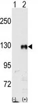
WB analysis of 293 cell lysate (2 ug/lane) either nontransfected (Lane 1) or transiently transfected with the EphB1 (Lane 2) using GTX81349 EphB1 antibody.
WB analysis of 293 cell lysate (2 ug/lane) either nontransfected (Lane 1) or transiently transfected with the EphB1 (Lane 2) using GTX81349 EphB1 antibody.
EphB1 antibody
GTX81349
ApplicationsWestern Blot
Product group Antibodies
ReactivityHuman
TargetEPHB1
Overview
- SupplierGeneTex
- Product NameEphB1 antibody
- Delivery Days Customer9
- Application Supplier NoteWB: 1:1000. *Optimal dilutions/concentrations should be determined by the researcher.Not tested in other applications.
- ApplicationsWestern Blot
- CertificationResearch Use Only
- ClonalityPolyclonal
- ConjugateUnconjugated
- Gene ID2047
- Target nameEPHB1
- Target descriptionEPH receptor B1
- Target synonymsELK, EPHT2, Hek6, NET, ephrin type-B receptor 1, EK6, EPH-like kinase 6, eph tyrosine kinase 2, neuronally-expressed EPH-related tyrosine kinase, tyrosine-protein kinase receptor EPH-2
- HostRabbit
- IsotypeIgG
- Protein IDP54762
- Protein NameEphrin type-B receptor 1
- Scientific DescriptionEphrin receptors and their ligands, the ephrins, mediate numerous developmental processes, particularly in the nervous system. Based on their structures and sequence relationships, ephrins are divided into the ephrin-A (EFNA) class, which are anchored to the membrane by a glycosylphosphatidylinositol linkage, and the ephrin-B (EFNB) class, which are transmembrane proteins. The Eph family of receptors are divided into 2 groups based on the similarity of their extracellular domain sequences and their affinities for binding ephrin-A and ephrin-B ligands. Ephrin receptors make up the largest subgroup of the receptor tyrosine kinase (RTK) family. The protein encoded by this gene is a receptor for ephrin-B family members. [provided by RefSeq, Jul 2008]
- ReactivityHuman
- Storage Instruction-20°C or -80°C,2°C to 8°C
- UNSPSC41116161
Datasheet
Related products
Product group Antibodies
Anti-EPHB1 AntibodyA97558
ApplicationsWestern Blot, ELISA
ReactivityHuman, Mouse, Rat
- SizePrice
Product group Antibodies
Anti-Ephb1 (Center) Antibody102-24089
ApplicationsWestern Blot
TargetEPHB1
- SizePrice
Product group Antibodies
ApplicationsFlow Cytometry, Western Blot, ImmunoCytoChemistry
ReactivityHuman, Mouse
TargetEPHB1
- SizePrice
Product group Antibodies
EPHB1/EPHB2 AntibodyCSB-PA002364
ApplicationsImmunoFluorescence, Western Blot, ELISA, ImmunoHistoChemistry
ReactivityHuman, Mouse, Rat
TargetEPHB1
- SizePrice
Product group Antibodies

ApplicationsWestern Blot
ReactivityHuman, Monkey, Mouse, Rat
TargetEPHB1
- SizePrice
![IHC-P analysis of human lung cancer (left) and colon cancer (right) using GTX83049 EphB1 antibody [5F10A4].](https://www.genetex.com/upload/website/prouct_img/normal/GTX83049/GTX83049_20170912_IHC-P_w_23061322_423.webp)
Product group Antibodies
EphB1 antibody [5F10A4]GTX83049
ApplicationsWestern Blot, ELISA, ImmunoHistoChemistry, ImmunoHistoChemistry Paraffin
ReactivityHuman
TargetEPHB1
- SizePrice

Product group Antibodies
Anti-Eph receptor B1/EPHB1 Antibody Picoband(r)PB9584-CARRIER-FREE
ApplicationsFlow Cytometry, Western Blot, ImmunoHistoChemistry
ReactivityHamster, Human, Mouse, Rat
TargetEPHB1
- SizePrice