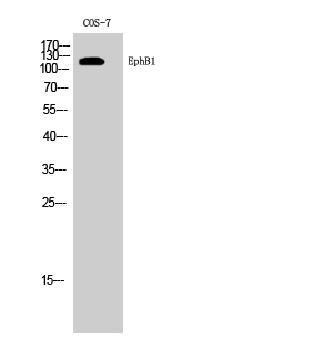
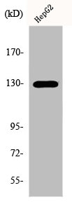
Bio-Connect
Anti-EPHB1 Antibody

Anti-EPHB1 Antibody

A97558
Antibodies.com
ApplicationsWestern Blot, ELISA
Product group Antibodies
Sign in to order and to see your custom pricing.
Large volume orders?
Order with a bulk request

Overview

  • Supplier
    Antibodies.com
  • Product Name
    Anti-EPHB1 Antibody
  • Delivery Days Customer
    7
  • Applications
    Western Blot, ELISA
  • Certification
    Research Use Only
  • Clonality
    Polyclonal
  • Conjugate
    Unconjugated
  • Host
    Rabbit
  • Isotype
    IgG
  • Scientific Description
    Rabbit polyclonal antibody to EPHB1.
  • UNSPSC
    12352203

Related products

Product group Antibodies
Assay Genie
TargetEPHB1
  • SizePrice
WB analysis of 293 cell lysate (2 ug/lane) either nontransfected (Lane 1) or transiently transfected with the EphB1 (Lane 2) using GTX81349 EphB1 antibody.
Product group Antibodies
GeneTex
ApplicationsWestern Blot
TargetEPHB1
  • SizePrice
Product group Antibodies
MedChem Express
TargetEPHB1
  • SizePrice
Western Blot analysis of HepG2 cells using EphB1/2 Polyclonal Antibody
Product group Antibodies
Cusabio
ApplicationsImmunoFluorescence, Western Blot, ELISA, ImmunoHistoChemistry
TargetEPHB1
  • SizePrice
Product group Antibodies
RayBiotech
ApplicationsWestern Blot
TargetEPHB1
  • SizePrice
Product group Antibodies
LifeSpan BioSciences
ApplicationsWestern Blot
TargetEPHB1
  • SizePrice
Product group Antibodies
Nordic-MUbio
ApplicationsImmunoPrecipitation, Western Blot
TargetEPHB1
  • SizePrice
Product group Antibodies
Bioss Antibodies
ApplicationsFlow Cytometry, Western Blot, ImmunoCytoChemistry
TargetEPHB1
  • SizePrice
Figure 1. Western blot analysis of Eph receptor B1 using anti-Eph receptor B1 antibody (PB9584). Electrophoresis was performed on a 5-20% SDS-PAGE gel at 70V (Stacking gel) / 90V (Resolving gel) for 2-3 hours. The sample well of each lane was loaded with 30 ug of sample under reducing conditions. Lane 1: human U251 whole cell lysates, Lane 2: human Hel whole cell lysates, Lane 3: rat testis tissue lysates, Lane 4: rat brain tissue lysates, Lane 5: mouse testis tissue lysates, Lane 6: mouse brain tissue lysates. After electrophoresis, proteins were transferred to a nitrocellulose membrane at 150 mA for 50-90 minutes. Blocked the membrane with 5% non-fat milk/TBS for 1.5 hour at RT. The membrane was incubated with rabbit anti-Eph receptor B1 antigen affinity purified polyclonal antibody (Catalog # PB9584) at 0.5 microg/mL overnight at 4°C, then washed with TBS-0.1%Tween 3 times with 5 minutes each and probed with a goat anti-rabbit IgG-HRP secondary antibody at a dilution of 1:5000 for 1.5 hour at RT. The signal is developed using an Enhanced Chemiluminescent detection (ECL) kit (Catalog # EK1002) with Tanon 5200 system. A specific band was detected for Eph receptor B1 at approximately 120 kDa. The expected band size for Eph receptor B1 is at 110 kDa.
Product group Antibodies
Boster Bio
ApplicationsFlow Cytometry, Western Blot, ImmunoHistoChemistry
TargetEPHB1
  • SizePrice