


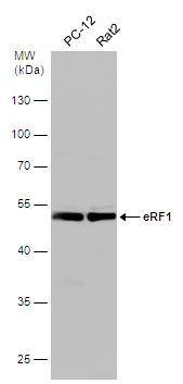
Figure 1. Western blot analysis of ETF1 using anti-ETF1 antibody (A04157-2). Electrophoresis was performed on a 5-20% SDS-PAGE gel at 70V (Stacking gel) / 90V (Resolving gel) for 2-3 hours. The sample well of each lane was loaded with 50ug of sample under reducing conditions. Lane 1: human Raji whole cell lysates, Lane 2: rat kidney tissue lysates, Lane 3: mouse NIH3T3 whole cell lysates, Lane 4: mouse RAW246.7 whole cell lysates. After Electrophoresis, proteins were transferred to a Nitrocellulose membrane at 150mA for 50-90 minutes. Blocked the membrane with 5% Non-fat Milk/ TBS for 1.5 hour at RT. The membrane was incubated with rabbit anti-ETF1 antigen affinity purified polyclonal antibody (Catalog # A04157-2) at 0.5 microg/mL overnight at 4°C, then washed with TBS-0.1%Tween 3 times with 5 minutes each and probed with a goat anti-rabbit IgG-HRP secondary antibody at a dilution of 1:5000 for 1.5 hour at RT. The signal is developed using an Enhanced Chemiluminescent detection (ECL) kit (Catalog # EK1002) with Tanon 5200 system. A specific band was detected for ETF1 at approximately 49KD. The expected band size for ETF1 is at 49KD.
Figure 1. Western blot analysis of ETF1 using anti-ETF1 antibody (A04157-2). Electrophoresis was performed on a 5-20% SDS-PAGE gel at 70V (Stacking gel) / 90V (Resolving gel) for 2-3 hours. The sample well of each lane was loaded with 50ug of sample under reducing conditions. Lane 1: human Raji whole cell lysates, Lane 2: rat kidney tissue lysates, Lane 3: mouse NIH3T3 whole cell lysates, Lane 4: mouse RAW246.7 whole cell lysates. After Electrophoresis, proteins were transferred to a Nitrocellulose membrane at 150mA for 50-90 minutes. Blocked the membrane with 5% Non-fat Milk/ TBS for 1.5 hour at RT. The membrane was incubated with rabbit anti-ETF1 antigen affinity purified polyclonal antibody (Catalog # A04157-2) at 0.5 microg/mL overnight at 4°C, then washed with TBS-0.1%Tween 3 times with 5 minutes each and probed with a goat anti-rabbit IgG-HRP secondary antibody at a dilution of 1:5000 for 1.5 hour at RT. The signal is developed using an Enhanced Chemiluminescent detection (ECL) kit (Catalog # EK1002) with Tanon 5200 system. A specific band was detected for ETF1 at approximately 49KD. The expected band size for ETF1 is at 49KD.
Anti-eRF1/ETF1 Picoband(r) Antibody
A04157-2-CARRIER-FREE
ApplicationsWestern Blot, ELISA
Product group Antibodies
ReactivityHuman, Mouse, Rat
TargetETF1
Overview
- SupplierBoster Bio
- Product NameAnti-eRF1/ETF1 Picoband(r) Antibody
- Delivery Days Customer9
- ApplicationsWestern Blot, ELISA
- CertificationResearch Use Only
- ClonalityPolyclonal
- Concentration500 ug/ml
- Gene ID2107
- Target nameETF1
- Target descriptioneukaryotic translation termination factor 1
- Target synonymsD5S1995, ERF, ERF1, RF1, SUP45L1, TB3-1, eukaryotic peptide chain release factor subunit 1, polypeptide chain release factor 1, protein Cl1, sup45 (yeast omnipotent suppressor 45) homolog-like 1
- HostRabbit
- IsotypeIgG
- Protein IDP62495
- Protein NameEukaryotic peptide chain release factor subunit 1
- Scientific DescriptionBoster Bio Anti-eRF1/ETF1 Picoband® Antibody catalog # A04157-2. Tested in ELISA, WB applications. This antibody reacts with Human, Mouse, Rat. The brand Picoband indicates this is a premium antibody that guarantees superior quality, high affinity, and strong signals with minimal background in Western blot applications. Only our best-performing antibodies are designated as Picoband, ensuring unmatched performance.
- ReactivityHuman, Mouse, Rat
- Storage Instruction-20°C,2°C to 8°C
- UNSPSC12352203
Related products
Product group Antibodies
ETF1 AntibodyCSB-PA007840GA01HU
ApplicationsWestern Blot, ELISA, ImmunoHistoChemistry
ReactivityHuman, Mouse, Rat
TargetETF1
- SizePrice
Product group Antibodies
Anti-ETF1 AntibodyA48548
ApplicationsWestern Blot, ELISA, ImmunoHistoChemistry
- SizePrice
Product group Antibodies
Anti-ETF1 AntibodyHPA037511
ApplicationsWestern Blot, ImmunoHistoChemistry
ReactivityHuman
TargetETF1
- SizePrice
Product group Antibodies

ETF1 / ERF1 AntibodyLS-C334382
ApplicationsWestern Blot
ReactivityHuman, Mouse, Rat
TargetETF1
- SizePrice
Product group Antibodies
eRF1 antibodyGTX108271
ApplicationsImmunoFluorescence, Western Blot, ImmunoCytoChemistry, ImmunoHistoChemistry, ImmunoHistoChemistry Paraffin
ReactivityHuman, Mouse, Rat
TargetETF1
- SizePrice
Product group Antibodies
Anti-ETF1Y158178
ApplicationsWestern Blot, ELISA, ImmunoHistoChemistry
ReactivityHuman, Mouse, Rat
- SizePrice
Product group Antibodies
Anti-ETF1 Antibody144-05920
ApplicationsWestern Blot
ReactivityHuman, Mouse, Rat
TargetETF1
- SizePrice
Product group Antibodies
eRF1 Polyclonal AntibodyBS-13098R
ApplicationsImmunoFluorescence, ELISA, ImmunoCytoChemistry, ImmunoHistoChemistry, ImmunoHistoChemistry Frozen, ImmunoHistoChemistry Paraffin
ReactivityBovine, Canine, Equine, Human, Mouse, Plant, Rat, Xenopus, Zebra Fish
TargetETF1
- SizePrice