

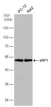
Various whole cell extracts (30 μg) were separated by 10% SDS-PAGE, and the membrane was blotted with eRF1 antibody (GTX108271) diluted at 1:500.
 diluted at 1:500.
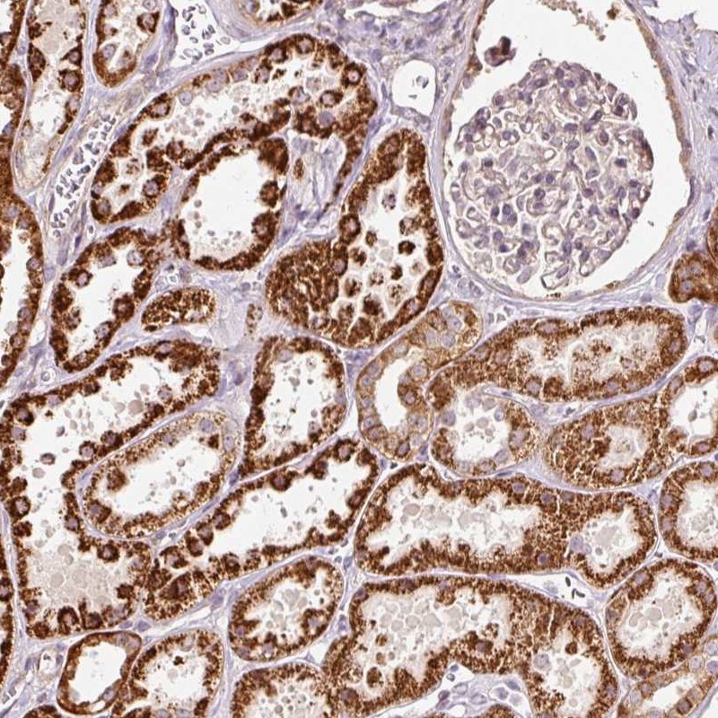
Antigen Retrieval: Trilogy? (EDTA based, pH 8.0) buffer, 15min")
 diluted at 1:500.
Antigen Retrieval: Trilogy? (EDTA based, pH 8.0) buffer, 15min")
![eRF1 antibody detects eRF1 protein at cytoplasm by immunofluorescent analysis. Sample: A431 cells were fixed in 4% paraformaldehyde at RT for 15 min. Green: eRF1 protein stained by eRF1 antibody (GTX108271) diluted at 1:500. Red: alpha Tubulin, a cytoskeleton marker, stained by alpha Tubulin antibody [GT114] (GTX628802) diluted at 1:1000. Blue: Hoechst 33342 staining.](https://www.genetex.com/upload/website/prouct_img/normal/GTX108271/GTX108271_39778_20150410_IFA_2_w_23060120_449.webp "eRF1 antibody detects eRF1 protein at cytoplasm by immunofluorescent analysis. Sample: A431 cells were fixed in 4% paraformaldehyde at RT for 15 min. Green: eRF1 protein stained by eRF1 antibody (GTX108271) diluted at 1:500. Red: alpha Tubulin, a cytoskeleton marker, stained by alpha Tubulin antibody [GT114] (GTX628802) diluted at 1:1000. Blue: Hoechst 33342 staining.")
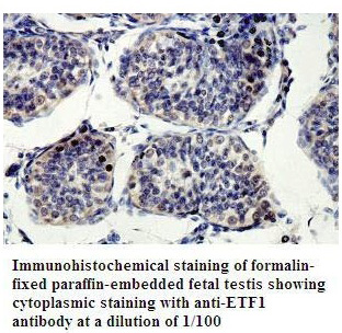
 antibody at 1:100 dilution.
Antigen Retrieval: Trilogy? (EDTA based, pH 8.0) buffer, 15min")
 were separated by 10% SDS-PAGE, and the membrane was blotted with eRF1 antibody (GTX108271) diluted at 1:500.")
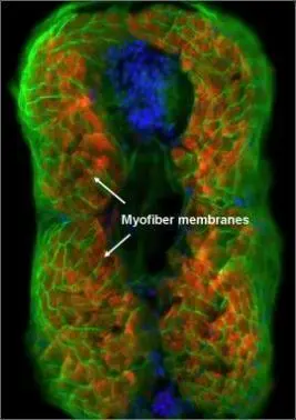
 diluted at 1:500. Red: phalloidin, a cytoskeleton marker, stained by phalloidin (invitrogen, A12380) diluted at 1:200. Blue: Hoechst 33342 staining.")
Various whole cell extracts (30 μg) were separated by 10% SDS-PAGE, and the membrane was blotted with eRF1 antibody (GTX108271) diluted at 1:500.
eRF1 antibody
GTX108271
ApplicationsImmunoFluorescence, Western Blot, ImmunoCytoChemistry, ImmunoHistoChemistry, ImmunoHistoChemistry Paraffin
Product group Antibodies
ReactivityHuman, Mouse, Rat
TargetETF1
Overview
- SupplierGeneTex
- Product NameeRF1 antibody
- Delivery Days Customer9
- Application Supplier NoteWB: 1:500-1:3000. ICC/IF: 1:100-1:1000. IHC-P: 1:100-1:1000. *Optimal dilutions/concentrations should be determined by the researcher.Not tested in other applications.
- ApplicationsImmunoFluorescence, Western Blot, ImmunoCytoChemistry, ImmunoHistoChemistry, ImmunoHistoChemistry Paraffin
- CertificationResearch Use Only
- ClonalityPolyclonal
- Concentration0.91 mg/ml
- ConjugateUnconjugated
- Gene ID2107
- Target nameETF1
- Target descriptioneukaryotic translation termination factor 1
- Target synonymsD5S1995, ERF, ERF1, RF1, SUP45L1, TB3-1, eukaryotic peptide chain release factor subunit 1, polypeptide chain release factor 1, protein Cl1, sup45 (yeast omnipotent suppressor 45) homolog-like 1
- HostRabbit
- IsotypeIgG
- Protein IDP62495
- Protein NameEukaryotic peptide chain release factor subunit 1
- Scientific DescriptionTermination of protein biosynthesis and release of the nascent polypeptide chain are signaled by the presence of an in-frame stop codon at the aminoacyl site of the ribosome. The process of translation termination is universal and is mediated by protein release factors (RFs) and GTP. A class 1 RF recognizes the stop codon and promotes the hydrolysis of the ester bond linking the polypeptide chain with the peptidyl site tRNA, a reaction catalyzed at the peptidyl transferase center of the ribosome. Class 2 RFs, which are not codon specific and do not recognize codons, stimulate class 1 RF activity and confer GTP dependency upon the process. In prokaryotes, both class 1 RFs, RF1 and RF2, recognize UAA; however, UAG and UGA are decoded specifically by RF1 and RF2, respectively. In eukaryotes, eRF1, or ETF1, the functional counterpart of RF1 and RF2, functions as an omnipotent RF, decoding all 3 stop codons (Frolova et al., 1994 [PubMed 7990965]).[supplied by OMIM]
- ReactivityHuman, Mouse, Rat
- Storage Instruction-20°C or -80°C,2°C to 8°C
- UNSPSC41116161
Datasheet
Related products
Product group Antibodies
ETF1 AntibodyCSB-PA007840GA01HU
ApplicationsWestern Blot, ELISA, ImmunoHistoChemistry
ReactivityHuman, Mouse, Rat
TargetETF1
- SizePrice
Product group Antibodies
Anti-ETF1 AntibodyA48548
ApplicationsWestern Blot, ELISA, ImmunoHistoChemistry
- SizePrice
Product group Antibodies
Anti-ETF1 AntibodyHPA037511
ApplicationsWestern Blot, ImmunoHistoChemistry
ReactivityHuman
TargetETF1
- SizePrice

Product group Antibodies
Anti-eRF1/ETF1 Picoband(r) AntibodyA04157-2-CARRIER-FREE
ApplicationsWestern Blot, ELISA
ReactivityHuman, Mouse, Rat
TargetETF1
- SizePrice
Product group Antibodies

ETF1 / ERF1 AntibodyLS-C334382
ApplicationsWestern Blot
ReactivityHuman, Mouse, Rat
TargetETF1
- SizePrice
Product group Antibodies
eRF1 antibodyGTX108296
ApplicationsImmunoFluorescence, Western Blot, ImmunoCytoChemistry, ImmunoHistoChemistry, ImmunoHistoChemistry Paraffin
ReactivityHuman, Mouse, Zebra Fish
TargetETF1
- SizePrice
Product group Antibodies
Anti-ETF1Y158178
ApplicationsWestern Blot, ELISA, ImmunoHistoChemistry
ReactivityHuman, Mouse, Rat
- SizePrice
Product group Antibodies
Anti-ETF1 Antibody144-05920
ApplicationsWestern Blot
ReactivityHuman, Mouse, Rat
TargetETF1
- SizePrice