


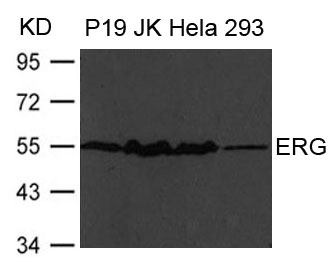
Figure 1. Western blot analysis of ERG using anti-ERG antibody (A00793-1). Electrophoresis was performed on a 5-20% SDS-PAGE gel at 70V (Stacking gel) / 90V (Resolving gel) for 2-3 hours. The sample well of each lane was loaded with 30 ug of sample under reducing conditions. Lane 1: human Colo320 whole cell lysates, Lane 2: rat liver tissue lysates, Lane 3: mouse heart tissue lysates, Lane 4: mouse kidney tissue lysates. After electrophoresis, proteins were transferred to a nitrocellulose membrane at 150 mA for 50-90 minutes. Blocked the membrane with 5% non-fat milk/TBS for 1.5 hour at RT. The membrane was incubated with rabbit anti-ERG antigen affinity purified polyclonal antibody (Catalog # A00793-1) at 0.5 microg/mL overnight at 4°C, then washed with TBS-0.1%Tween 3 times with 5 minutes each and probed with a goat anti-rabbit IgG-HRP secondary antibody at a dilution of 1:5000 for 1.5 hour at RT. The signal is developed using an Enhanced Chemiluminescent detection (ECL) kit (Catalog # EK1002) with Tanon 5200 system. A specific band was detected for ERG at approximately 55 kDa. The expected band size for ERG is at 55 kDa.
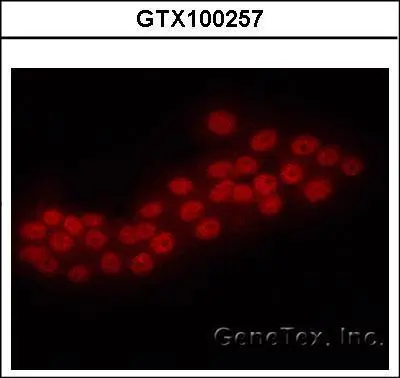
. ERG was detected in an immunocytochemical section of A431 cells. Enzyme antigen retrieval was performed using IHC enzyme antigen retrieval reagent (AR0022) for 15 mins. The cells were blocked with 10% goat serum. And then incubated with 5 microg/mL rabbit anti-ERG Antibody (A00793-1) overnight at 4°C. DyLight®488 Conjugated Goat Anti-Rabbit IgG (BA1127) was used as secondary antibody at 1:100 dilution and incubated for 30 minutes at 37°C. The section was counterstained with DAPI. Visualize using a fluorescence microscope and filter sets appropriate for the label used.")
Figure 1. Western blot analysis of ERG using anti-ERG antibody (A00793-1). Electrophoresis was performed on a 5-20% SDS-PAGE gel at 70V (Stacking gel) / 90V (Resolving gel) for 2-3 hours. The sample well of each lane was loaded with 30 ug of sample under reducing conditions. Lane 1: human Colo320 whole cell lysates, Lane 2: rat liver tissue lysates, Lane 3: mouse heart tissue lysates, Lane 4: mouse kidney tissue lysates. After electrophoresis, proteins were transferred to a nitrocellulose membrane at 150 mA for 50-90 minutes. Blocked the membrane with 5% non-fat milk/TBS for 1.5 hour at RT. The membrane was incubated with rabbit anti-ERG antigen affinity purified polyclonal antibody (Catalog # A00793-1) at 0.5 microg/mL overnight at 4°C, then washed with TBS-0.1%Tween 3 times with 5 minutes each and probed with a goat anti-rabbit IgG-HRP secondary antibody at a dilution of 1:5000 for 1.5 hour at RT. The signal is developed using an Enhanced Chemiluminescent detection (ECL) kit (Catalog # EK1002) with Tanon 5200 system. A specific band was detected for ERG at approximately 55 kDa. The expected band size for ERG is at 55 kDa.
Anti-ERG Antibody Picoband(r)
A00793-1-CARRIER-FREE
ApplicationsImmunoFluorescence, Western Blot, ELISA, ImmunoCytoChemistry
Product group Antibodies
ReactivityHuman, Mouse, Rat
TargetERG
Overview
- SupplierBoster Bio
- Product NameAnti-ERG Antibody Picoband(r)
- Delivery Days Customer9
- ApplicationsImmunoFluorescence, Western Blot, ELISA, ImmunoCytoChemistry
- CertificationResearch Use Only
- ClonalityPolyclonal
- Concentration500 ug/ml
- Gene ID2078
- Target nameERG
- Target descriptionETS transcription factor ERG
- Target synonymsLMPHM14, erg-3, p55, transcriptional regulator ERG, ERG, ETS transcription factor, FUS/ERG fusion protein, TMPRSS2/ERG fusion, ets-related, transcriptional regulator ERG (transforming protein ERG), v-ets avian erythroblastosis virus E26 oncogene homolog, v-ets avian erythroblastosis virus E26 oncogene related, v-ets erythroblastosis virus E26 oncogene homolog, v-ets erythroblastosis virus E26 oncogene like
- HostRabbit
- IsotypeIgG
- Protein IDP11308
- Protein NameTranscriptional regulator ERG
- Scientific DescriptionBoster Bio Anti-ERG Antibody Picoband® catalog # A00793-1. Tested in ELISA, IF, ICC, WB applications. This antibody reacts with Human, Mouse, Rat. The brand Picoband indicates this is a premium antibody that guarantees superior quality, high affinity, and strong signals with minimal background in Western blot applications. Only our best-performing antibodies are designated as Picoband, ensuring unmatched performance.
- ReactivityHuman, Mouse, Rat
- Storage Instruction-20°C,2°C to 8°C
- UNSPSC12352203
Related products
Product group Antibodies
Anti-ERG AntibodyA44230
ApplicationsWestern Blot
ReactivityHuman, Mouse, Rat
- SizePrice
Product group Antibodies
Anti-ERG Antibody144-01240
ApplicationsImmunoFluorescence, Western Blot
ReactivityHuman, Mouse
TargetERG
- SizePrice

Product group Antibodies
ERG Recombinant AntibodyBSM-52324R
ApplicationsFlow Cytometry, ImmunoFluorescence, Western Blot, ImmunoCytoChemistry, ImmunoHistoChemistry, ImmunoHistoChemistry Frozen, ImmunoHistoChemistry Paraffin
ReactivityHuman, Mouse, Rat
TargetERG
- SizePrice
Product group Antibodies
ERG AntibodyCSB-PA007781HA01HU
ApplicationsImmunoFluorescence, ImmunoPrecipitation, ELISA, ImmunoHistoChemistry
ReactivityHuman, Rat
TargetERG
- SizePrice
Product group Antibodies

Goat anti-ERGEB07081
ApplicationsFlow Cytometry, ImmunoFluorescence, Western Blot, ELISA
ReactivityHuman
TargetERG
- SizePrice
Product group Antibodies
Erg Polyclonal AntibodyCAC11844
ApplicationsImmunoFluorescence, ImmunoPrecipitation, ELISA, ImmunoHistoChemistry
ReactivityRat
TargetERG
- SizePrice
Product group Antibodies

ERG AntibodyLS-C400782
ApplicationsELISA, ImmunoHistoChemistry
ReactivityHuman, Mouse
TargetERG
- SizePrice
Product group Antibodies
ERG antibody [C3], C-termGTX100257
ApplicationsImmunoFluorescence, Western Blot, ImmunoCytoChemistry
ReactivityHuman, Mouse
TargetERG
- SizePrice

Product group Antibodies
Anti-ERG AntibodyHPA046598
ApplicationsChIP Chromatin ImmunoPrecipitation, ImmunoHistoChemistry
ReactivityHuman
TargetERG
- SizePrice