

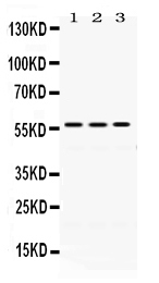
Figure 1. Western blot analysis of ERp57 using anti-ERp57 antibody (PB9772). Electrophoresis was performed on a 5-20% SDS-PAGE gel at 70V (Stacking gel) / 90V (Resolving gel) for 2-3 hours. The sample well of each lane was loaded with 50ug of sample under reducing conditions. Lane 1: Rat Liver Tissue Lysate, Lane 2: Mouse Liver Tissue Lysate, Lane 3: A549 Whole Cell Lysate. After Electrophoresis, proteins were transferred to a Nitrocellulose membrane at 150mA for 50-90 minutes. Blocked the membrane with 5% Non-fat Milk/ TBS for 1.5 hour at RT. The membrane was incubated with rabbit anti-ERp57 antigen affinity purified polyclonal antibody (Catalog # PB9772) at 0.5 microg/mL overnight at 4°C, then washed with TBS-0.1%Tween 3 times with 5 minutes each and probed with a goat anti-rabbit IgG-HRP secondary antibody at a dilution of 1:10000 for 1.5 hour at RT. The signal is developed using an Enhanced Chemiluminescent detection (ECL) kit (Catalog # EK1002) with Tanon 5200 system. A specific band was detected for ERp57 at approximately 57KD. The expected band size for ERp57 is at 57KD.
. PB9771 was detected in paraffin-embedded section of Human Lung Cancer Tissue. Heat mediated antigen retrieval was performed in citrate buffer (pH6, epitope retrieval solution) for 20 mins. The tissue section was blocked with 10% goat serum. The tissue section was then incubated with 1microg/ml rabbit anti-PB9771 Antibody (PB9772) overnight at 4°C. Biotinylated goat anti-rabbit IgG was used as secondary antibody and incubated for 30 minutes at 37°C. The tissue section was developed using Strepavidin-Biotin-Complex (SABC)(Catalog # SA1022) with DAB as the chromogen.")
. ERp57 was detected in frozen section of human placenta tissue . The tissue section was blocked with 10% goat serum. The tissue section was then incubated with 1microg/ml rabbit anti-ERp57 Antibody (PB9772) overnight at 4°C. Biotinylated goat anti-rabbit IgG was used as secondary antibody and incubated for 30 minutes at 37°C. The tissue section was developed using Strepavidin-Biotin-Complex (SABC)(Catalog # SA1022) with DAB as the chromogen.")
. ERp57 was detected in immunocytochemical section of U20S cells. Enzyme antigen retrieval was performed using IHC enzyme antigen retrieval reagent (AR0022) for 15 mins. The cells were blocked with 10% goat serum. And then incubated with 2microg/mL rabbit anti-ERp57 Antibody (PB9772) overnight at 4°C. DyLight®488 Conjugated Goat Anti-Rabbit IgG (BA1127) was used as secondary antibody at 1:100 dilution and incubated for 30 minutes at 37°C. The section was counterstained with DAPI. Visualize using a fluorescence microscope and filter sets appropriate for the label used.")
. Overlay histogram showing PC-3 cells stained with PB9772 (Blue line). To facilitate intracellular staining, cells were fixed with 4% paraformaldehyde and permeabilized with permeabilization buffer. The cells were blocked with 10% normal goat serum. And then incubated with rabbit anti-ERp57 Antibody (PB9772, 1microg/1x106 cells) for 30 min at 20°C. DyLight®488 conjugated goat anti-rabbit IgG (BA1127, 5-10microg/1x106 cells) was used as secondary antibody for 30 minutes at 20°C. Isotype control antibody (Green line) was rabbit IgG (1microg/1x106) used under the same conditions. Unlabelled sample without incubation with primary antibody and secondary antibody (Red line) was used as a blank control.")
Figure 1. Western blot analysis of ERp57 using anti-ERp57 antibody (PB9772). Electrophoresis was performed on a 5-20% SDS-PAGE gel at 70V (Stacking gel) / 90V (Resolving gel) for 2-3 hours. The sample well of each lane was loaded with 50ug of sample under reducing conditions. Lane 1: Rat Liver Tissue Lysate, Lane 2: Mouse Liver Tissue Lysate, Lane 3: A549 Whole Cell Lysate. After Electrophoresis, proteins were transferred to a Nitrocellulose membrane at 150mA for 50-90 minutes. Blocked the membrane with 5% Non-fat Milk/ TBS for 1.5 hour at RT. The membrane was incubated with rabbit anti-ERp57 antigen affinity purified polyclonal antibody (Catalog # PB9772) at 0.5 microg/mL overnight at 4°C, then washed with TBS-0.1%Tween 3 times with 5 minutes each and probed with a goat anti-rabbit IgG-HRP secondary antibody at a dilution of 1:10000 for 1.5 hour at RT. The signal is developed using an Enhanced Chemiluminescent detection (ECL) kit (Catalog # EK1002) with Tanon 5200 system. A specific band was detected for ERp57 at approximately 57KD. The expected band size for ERp57 is at 57KD.
Anti-ERp57/PDIA3 Antibody Picoband(r)
PB9772-CARRIER-FREE
ApplicationsFlow Cytometry, ImmunoFluorescence, Western Blot, ImmunoCytoChemistry, ImmunoHistoChemistry, ImmunoHistoChemistry Frozen
Product group Antibodies
ReactivityBovine, Human, Mouse, Rat
TargetPDIA3
Overview
- SupplierBoster Bio
- Product NameAnti-ERp57/PDIA3 Antibody Picoband(r)
- Delivery Days Customer9
- Application Supplier NoteTested Species: In-house tested species with positive results. By Heat: Boiling the paraffin sections in 10mM citrate buffer, pH6.0, for 20mins is required for the staining of formalin/paraffin sections. Other applications have not been tested. Optimal dilutions should be determined by end users.
- ApplicationsFlow Cytometry, ImmunoFluorescence, Western Blot, ImmunoCytoChemistry, ImmunoHistoChemistry, ImmunoHistoChemistry Frozen
- CertificationResearch Use Only
- ClonalityPolyclonal
- Concentration500 ug/ml
- Gene ID2923
- Target namePDIA3
- Target descriptionprotein disulfide isomerase family A member 3
- Target synonymsER60, ERp57, ERp60, ERp61, GRP57, GRP58, HEL-S-269, HEL-S-93n, HsT17083, P58, PI-PLC, protein disulfide-isomerase A3, 58 kDa glucose-regulated protein, 58 kDa microsomal protein, ER protein 57, ER protein 60, disulfide isomerase ER-60, endoplasmic reticulum P58, endoplasmic reticulum resident protein 57, endoplasmic reticulum resident protein 60, epididymis secretory protein Li 269, epididymis secretory sperm binding protein Li 93n, glucose regulated protein, 58kDa, phospholipase C-alpha, protein disulfide isomerase-associated 3
- HostRabbit
- IsotypeIgG
- Protein IDP30101
- Protein NameProtein disulfide-isomerase A3
- Scientific DescriptionBoster Bio Anti-ERp57/PDIA3 Antibody Picoband® catalog # PB9772. Tested in Flow Cytometry, IF, IHC, IHC-F, ICC, WB applications. This antibody reacts with Human, Mouse, Rat. The brand Picoband indicates this is a premium antibody that guarantees superior quality, high affinity, and strong signals with minimal background in Western blot applications. Only our best-performing antibodies are designated as Picoband, ensuring unmatched performance.
- ReactivityBovine, Human, Mouse, Rat
- Storage Instruction-20°C,2°C to 8°C
- UNSPSC12352203
Related products
Product group Antibodies
PDIA3 AntibodyCSB-PA005150
ApplicationsWestern Blot, ELISA
ReactivityHuman, Mouse, Rat
TargetPDIA3
- SizePrice

Product group Antibodies
Anti-PDIA3 AntibodyA97355
ApplicationsWestern Blot, ELISA
ReactivityHuman, Mouse, Rat
- SizePrice

Product group Antibodies
Anti-PDIA3 AntibodyAMAB90988
ApplicationsWestern Blot, ImmunoCytoChemistry, ImmunoHistoChemistry
ReactivityHuman
TargetPDIA3
- SizePrice
Product group Antibodies

ApplicationsWestern Blot, ELISA
ReactivityBovine, Human, Mouse, Rat
TargetPDIA3
- SizePrice
Product group Antibodies

PDIA3 / ERp57 AntibodyLS-C331251
ApplicationsWestern Blot, ImmunoHistoChemistry
ReactivityHuman, Mouse, Rat
TargetPDIA3
- SizePrice
Product group Antibodies
ApplicationsImmunoPrecipitation, Western Blot, ImmunoCytoChemistry, ImmunoHistoChemistry
ReactivityMouse, Porcine, Rat
TargetPDIA3
- SizePrice
![Various whole cell extracts (30 μg) were separated by 10% SDS-PAGE, and the membrane was blotted with ERp57 antibody [C3], C-term (GTX100297) diluted at 1:10000. The HRP-conjugated anti-rabbit IgG antibody (GTX213110-01) was used to detect the primary antibody.](https://www.genetex.com/upload/website/prouct_img/normal/GTX100297/GTX100297_39422_20190624_WB_R_w_23060100_776.webp)
Product group Antibodies
ERp57 antibody [C3], C-termGTX100297
ApplicationsImmunoFluorescence, Western Blot, ImmunoCytoChemistry, ImmunoHistoChemistry, ImmunoHistoChemistry Paraffin
ReactivityHuman, Mouse, Rat
TargetPDIA3
- SizePrice
Product group Antibodies
ERp57 Recombinant Antibody, AbBy Fluor-350 ConjugatedBSM-61463R-BF350
ApplicationsImmunoFluorescence, Western Blot
ReactivityHuman
TargetPDIA3
- SizePrice