


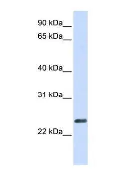
Figure 1. Western blot analysis of ERVW-1 using anti-ERVW-1 antibody (PB9622). Electrophoresis was performed on a 5-20% SDS-PAGE gel at 70V (Stacking gel) / 90V (Resolving gel) for 2-3 hours. The sample well of each lane was loaded with 30 ug of sample under reducing conditions. Lane 1: human Hela whole cell lysates, Lane 2: human K562 whole cell lysates, Lane 3: human SH-SY5Y whole cell lysates, Lane 4: human MCF-7 whole cell lysates. After electrophoresis, proteins were transferred to a nitrocellulose membrane at 150 mA for 50-90 minutes. Blocked the membrane with 5% non-fat milk/TBS for 1.5 hour at RT. The membrane was incubated with rabbit anti-ERVW-1 antigen affinity purified polyclonal antibody (Catalog # PB9622) at 0.5 microg/mL overnight at 4°C, then washed with TBS-0.1%Tween 3 times with 5 minutes each and probed with a goat anti-rabbit IgG-HRP secondary antibody at a dilution of 1:5000 for 1.5 hour at RT. The signal is developed using an Enhanced Chemiluminescent detection (ECL) kit (Catalog # EK1002) with Tanon 5200 system. A specific band was detected for ERVW-1 at approximately 80 kDa. The expected band size for ERVW-1 is at 80 kDa.
Figure 1. Western blot analysis of ERVW-1 using anti-ERVW-1 antibody (PB9622). Electrophoresis was performed on a 5-20% SDS-PAGE gel at 70V (Stacking gel) / 90V (Resolving gel) for 2-3 hours. The sample well of each lane was loaded with 30 ug of sample under reducing conditions. Lane 1: human Hela whole cell lysates, Lane 2: human K562 whole cell lysates, Lane 3: human SH-SY5Y whole cell lysates, Lane 4: human MCF-7 whole cell lysates. After electrophoresis, proteins were transferred to a nitrocellulose membrane at 150 mA for 50-90 minutes. Blocked the membrane with 5% non-fat milk/TBS for 1.5 hour at RT. The membrane was incubated with rabbit anti-ERVW-1 antigen affinity purified polyclonal antibody (Catalog # PB9622) at 0.5 microg/mL overnight at 4°C, then washed with TBS-0.1%Tween 3 times with 5 minutes each and probed with a goat anti-rabbit IgG-HRP secondary antibody at a dilution of 1:5000 for 1.5 hour at RT. The signal is developed using an Enhanced Chemiluminescent detection (ECL) kit (Catalog # EK1002) with Tanon 5200 system. A specific band was detected for ERVW-1 at approximately 80 kDa. The expected band size for ERVW-1 is at 80 kDa.
Anti-ERVW-1 Antibody Picoband(r)
PB9622
ApplicationsWestern Blot
Product group Antibodies
ReactivityHuman
TargetERVW-1
Overview
- SupplierBoster Bio
- Product NameAnti-ERVW-1 Antibody Picoband(r)
- Delivery Days Customer9
- Application Supplier NoteTested Species: In-house tested species with positive results. Other applications have not been tested. Optimal dilutions should be determined by end users.
- ApplicationsWestern Blot
- CertificationResearch Use Only
- ClonalityPolyclonal
- Concentration500 ug/ml
- Gene ID30816
- Target nameERVW-1
- Target descriptionendogenous retrovirus group W member 1, envelope
- Target synonymsENV, ENVW, ERVWE1, HERV-7q, HERV-W-ENV, HERV7Q, HERVW, HERVWENV, syncytin-1, HERV-7q envelope protein, HERV-W Env glycoprotein, HERV-W envelope protein, HERV-W_7q21.2 provirus ancestral Env polyprotein, HERV-tryptophan envelope protein, endogenous retroviral family W, env(C7), member 1, endogenous retrovirus group W, member 1, envelope glycoprotein, envelope polyprotein gPr73, enverin, human endogenous retrovirus W envC7-1 envelope protein
- HostRabbit
- IsotypeIgG
- Protein IDQ9UQF0
- Protein NameSyncytin-1
- Scientific DescriptionBoster Bio Anti-ERVW-1 Antibody Picoband® catalog # PB9622. Tested in WB applications. This antibody reacts with Human. The brand Picoband indicates this is a premium antibody that guarantees superior quality, high affinity, and strong signals with minimal background in Western blot applications. Only our best-performing antibodies are designated as Picoband, ensuring unmatched performance.
- ReactivityHuman
- Storage Instruction-20°C,2°C to 8°C
- UNSPSC12352203
Datasheet
MSDS
Related products
Product group Antibodies
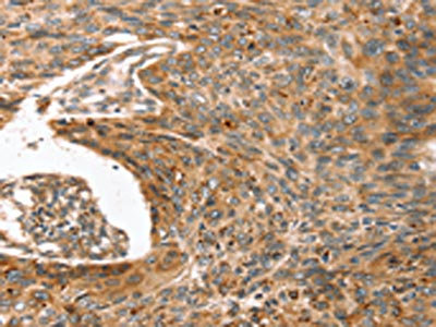
Anti-ERVW-1 AntibodyA45628
ApplicationsImmunoHistoChemistry
ReactivityHuman
- SizePrice
Product group Antibodies
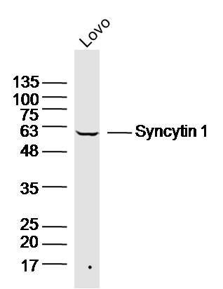
Anti-ERVW-1 Antibody144-64458
ApplicationsWestern Blot
ReactivityHuman, Mouse
TargetERVW-1
- SizePrice
Product group Antibodies
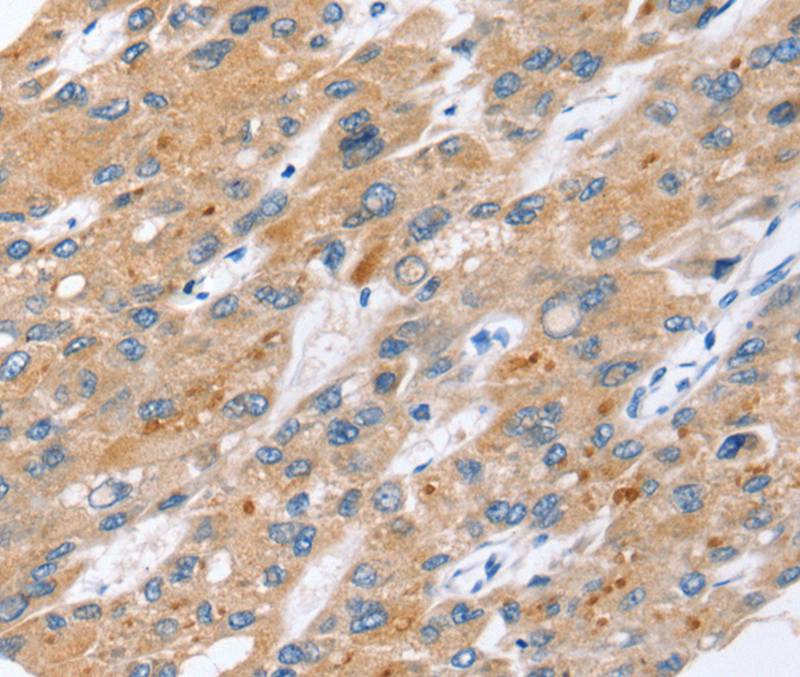
Ervw-1 Polyclonal AntibodyCAC09941
ApplicationsImmunoFluorescence, ELISA, ImmunoHistoChemistry
TargetERVW-1
- SizePrice
Product group Antibodies
ERVW-1 AntibodyCSB-PA215261
ApplicationsELISA, ImmunoHistoChemistry
ReactivityHuman
TargetERVW-1
- SizePrice
Product group Antibodies
References
Syncytin 1 Polyclonal AntibodyBS-2962R
ApplicationsWestern Blot, ELISA
ReactivityHuman, Mouse
TargetERVW-1
- SizePrice
Product group Antibodies

ApplicationsELISA, ImmunoHistoChemistry
ReactivityHuman
TargetERVW-1
- SizePrice
Product group Antibodies
References
ERVWE1 antibody, N-termGTX46373
ApplicationsWestern Blot
ReactivityHamster, Human
TargetERVW-1
- SizePrice
Product group Antibodies
Anti-ERVW-1 Antibody Picoband(r)PB9622-CARRIER-FREE
ApplicationsWestern Blot
ReactivityHuman
TargetERVW-1
- SizePrice