


Immunohistochemical staining of human spleen shows weak membranous positivity in cells in red pulp.
Immunohistochemical staining of human spleen shows weak membranous positivity in cells in red pulp.
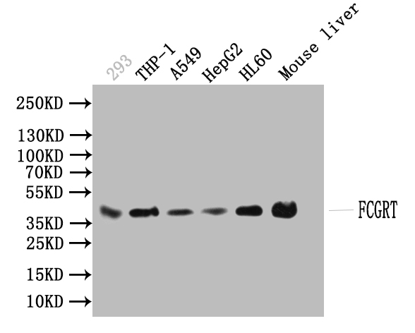
Anti-FCGRT Antibody
HPA015130
ApplicationsImmunoHistoChemistry
Product group Antibodies
ReactivityHuman
TargetFCGRT
Overview
- SupplierAtlas Antibodies
- Product NameAnti-FCGRT Antibody
- Delivery Days Customer4
- ApplicationsImmunoHistoChemistry
- CertificationResearch Use Only
- ClonalityPolyclonal
- ConjugateUnconjugated
- Gene ID2217
- Target nameFCGRT
- Target descriptionFc gamma receptor and transporter
- Target synonymsFCRN, FcgammaRn, alpha-chain, IgG receptor FcRn large subunit p51, Fc fragment of IgG receptor and transporter, Fc fragment of IgG, receptor, transporter, alpha, FcRn alpha chain, IgG Fc fragment receptor transporter alpha chain, heavy chain of the major histocompatibility complex class I-like Fc receptor, immunoglobulin receptor, intestinal, heavy chain, major histocompatibility complex class I-like Fc receptor, neonatal Fc receptor, neonatal Fc-receptor for Ig, neonatal fragment crystallizable Fc receptor FcRn, transmembrane alpha chain of the neonatal receptor
- HostRabbit
- IsotypeIgG
- Protein IDP55899
- Protein NameIgG receptor FcRn large subunit p51
- Scientific DescriptionRecombinant Protein Epitope Signature Tag (PrEST) antigen sequence
- ReactivityHuman
- Storage Instruction-20°C,2°C to 8°C
- UNSPSC41116161
Datasheet
MSDS
Related products
Product group Antibodies
FCGRT AntibodyCSB-PA008545EA01HU
ApplicationsImmunoFluorescence, Western Blot, ELISA, ImmunoHistoChemistry
ReactivityHuman, Mouse
TargetFCGRT
- SizePrice

Product group Antibodies
Anti-FCGRT AntibodyAMAB91199
ApplicationsImmunoHistoChemistry
ReactivityHuman
TargetFCGRT
- SizePrice
Product group Antibodies
Anti-FCGRT AntibodyAMAB91199
ApplicationsImmunoHistoChemistry
ReactivityHuman
TargetFCGRT
- SizePrice

Product group Antibodies
Anti-FCGRT AntibodyAMAB91200
ApplicationsWestern Blot, ImmunoHistoChemistry
ReactivityHuman
TargetFCGRT
- SizePrice
Product group Antibodies

Anti-FcRn [HL161-11G]AB02467-10.0-BT
ApplicationsFlow Cytometry, Other Application
ReactivityHuman, Monkey, Mouse, Rat
TargetFCGRT
- SizePrice
Product group Antibodies

FCGRT / FCRN AntibodyLS-C830070
ApplicationsWestern Blot, ELISA, ImmunoHistoChemistry
ReactivityHuman
TargetFCGRT
- SizePrice

Product group Antibodies
Anti-FCGRT AntibodyHPA012122
ApplicationsWestern Blot, ImmunoHistoChemistry
ReactivityHuman
TargetFCGRT
- SizePrice