


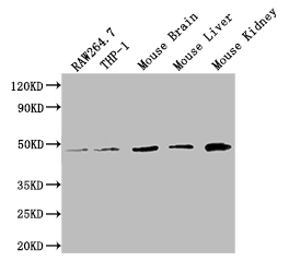
Figure 1. Western blot analysis of FENS1/WDFY1 using anti-FENS1/WDFY1 antibody (A10318-3). Electrophoresis was performed on a 5-20% SDS-PAGE gel at 70V (Stacking gel) / 90V (Resolving gel) for 2-3 hours. The sample well of each lane was loaded with 30 ug of sample under reducing conditions. Lane 1: human U-87MG whole cell lysates, Lane 2: human RT4 whole cell lysates, Lane 3: human THP-1 whole cell lysates, Lane 4: rat brain tissue lysates, Lane 5: rat skeletal muscle tissue lysates, Lane 6: mouse brain tissue lysates, Lane 7: mouse skeletal muscle tissue lysates. After electrophoresis, proteins were transferred to a nitrocellulose membrane at 150 mA for 50-90 minutes. Blocked the membrane with 5% non-fat milk/TBS for 1.5 hour at RT. The membrane was incubated with rabbit anti-FENS1/WDFY1 antigen affinity purified polyclonal antibody (Catalog # A10318-3) at 0.5 microg/mL overnight at 4°C, then washed with TBS-0.1%Tween 3 times with 5 minutes each and probed with a goat anti-rabbit IgG-HRP secondary antibody at a dilution of 1:5000 for 1.5 hour at RT. The signal is developed using an Enhanced Chemiluminescent detection (ECL) kit (Catalog # EK1002) with Tanon 5200 system. A specific band was detected for FENS1/WDFY1 at approximately 46 kDa. The expected band size for FENS1/WDFY1 is at 46 kDa.
. Overlay histogram showing HL-60 cells stained with A10318-3 (Blue line). To facilitate intracellular staining, cells were fixed with 4% paraformaldehyde and permeabilized with permeabilization buffer. The cells were blocked with 10% normal goat serum. And then incubated with rabbit anti-FENS1/WDFY1 Antibody (A10318-3, 1 microg/1x106 cells) for 30 min at 20°C. DyLight®488 conjugated goat anti-rabbit IgG (BA1127, 5-10 microg/1x106 cells) was used as secondary antibody for 30 minutes at 20°C. Isotype control antibody (Green line) was rabbit IgG (1 microg/1x106) used under the same conditions. Unlabelled sample without incubation with primary antibody and secondary antibody (Red line) was used as a blank control.")
Figure 1. Western blot analysis of FENS1/WDFY1 using anti-FENS1/WDFY1 antibody (A10318-3). Electrophoresis was performed on a 5-20% SDS-PAGE gel at 70V (Stacking gel) / 90V (Resolving gel) for 2-3 hours. The sample well of each lane was loaded with 30 ug of sample under reducing conditions. Lane 1: human U-87MG whole cell lysates, Lane 2: human RT4 whole cell lysates, Lane 3: human THP-1 whole cell lysates, Lane 4: rat brain tissue lysates, Lane 5: rat skeletal muscle tissue lysates, Lane 6: mouse brain tissue lysates, Lane 7: mouse skeletal muscle tissue lysates. After electrophoresis, proteins were transferred to a nitrocellulose membrane at 150 mA for 50-90 minutes. Blocked the membrane with 5% non-fat milk/TBS for 1.5 hour at RT. The membrane was incubated with rabbit anti-FENS1/WDFY1 antigen affinity purified polyclonal antibody (Catalog # A10318-3) at 0.5 microg/mL overnight at 4°C, then washed with TBS-0.1%Tween 3 times with 5 minutes each and probed with a goat anti-rabbit IgG-HRP secondary antibody at a dilution of 1:5000 for 1.5 hour at RT. The signal is developed using an Enhanced Chemiluminescent detection (ECL) kit (Catalog # EK1002) with Tanon 5200 system. A specific band was detected for FENS1/WDFY1 at approximately 46 kDa. The expected band size for FENS1/WDFY1 is at 46 kDa.
Anti-FENS1/WDFY1 Antibody Picoband(r)
A10318-3-CARRIER-FREE
ApplicationsFlow Cytometry, Western Blot, ELISA
Product group Antibodies
ReactivityHuman, Mouse, Rat
TargetWDFY1
Overview
- SupplierBoster Bio
- Product NameAnti-FENS1/WDFY1 Antibody Picoband(r)
- Delivery Days Customer9
- ApplicationsFlow Cytometry, Western Blot, ELISA
- CertificationResearch Use Only
- ClonalityPolyclonal
- Concentration500 ug/ml
- Gene ID57590
- Target nameWDFY1
- Target descriptionWD repeat and FYVE domain containing 1
- Target synonymsFENS-1, FENS1, WDF1, ZFYVE17, WD repeat and FYVE domain-containing protein 1, FYVE domain-containing protein localized to endosomes 1, WD40- and FYVE domain-containing protein 1, phosphoinositide-binding protein SR1, zinc finger FYVE domain-containing protein 17
- HostRabbit
- IsotypeIgG
- Protein IDQ8IWB7
- Protein NameWD repeat and FYVE domain-containing protein 1
- Scientific DescriptionBoster Bio Anti-FENS1/WDFY1 Antibody Picoband® catalog # A10318-3. Tested in ELISA, Flow Cytometry, WB applications. This antibody reacts with Human, Mouse, Rat. The brand Picoband indicates this is a premium antibody that guarantees superior quality, high affinity, and strong signals with minimal background in Western blot applications. Only our best-performing antibodies are designated as Picoband, ensuring unmatched performance.
- ReactivityHuman, Mouse, Rat
- Storage Instruction-20°C,2°C to 8°C
- UNSPSC12352203
Related products

Product group Antibodies

Goat anti-FENS1 / WDFY1EB06341
ApplicationsELISA
ReactivityHuman, Mouse, Rat
TargetWDFY1
- SizePrice

Product group Antibodies
Anti-WDFY1 AntibodyHPA050603
ApplicationsWestern Blot, ImmunoHistoChemistry
ReactivityHuman
TargetWDFY1
- SizePrice
Product group Antibodies
WDFY1 AntibodyCSB-PA816890LA01HU
ApplicationsWestern Blot, ELISA
ReactivityHuman, Mouse
TargetWDFY1
- SizePrice
Product group Antibodies

WDFY1 AntibodyLS-C501305
ApplicationsWestern Blot, ELISA
ReactivityHuman, Mouse
TargetWDFY1
- SizePrice
Product group Antibodies
Wdfy1 Polyclonal AntibodyCAC09470
ApplicationsWestern Blot, ELISA
ReactivityMouse
TargetWDFY1
- SizePrice
Product group Antibodies
WDFY1 AntibodyPACO49234
ApplicationsWestern Blot, ELISA
ReactivityHuman, Mouse
TargetWDFY1
- SizePrice
![Non-transfected (–) and transfected (+) 293T whole cell extracts (30 μg) were separated by 10% SDS-PAGE, and the membrane was blotted with WDFY1+WDFY2 antibody [HL1989] (GTX637890) diluted at 1:5000. The HRP-conjugated anti-rabbit IgG antibody (GTX213110-01) was used to detect the primary antibody.](https://www.genetex.com/upload/website/prouct_img/normal/GTX637890/GTX637890_T-44872_20240112_WB_multiple_B_24011618_608.webp)
Product group Antibodies
WDFY1 + WDFY2 antibody [HL1989]GTX637890
ApplicationsWestern Blot
ReactivityHuman
TargetWDFY1
- SizePrice
Product group Antibodies
FENS1 Polyclonal AntibodyBS-13169R
ApplicationsFlow Cytometry, ImmunoFluorescence, ELISA, ImmunoCytoChemistry, ImmunoHistoChemistry, ImmunoHistoChemistry Frozen, ImmunoHistoChemistry Paraffin
ReactivityBovine, Canine, Equine, Human, Mouse, Porcine, Rabbit, Rat
TargetWDFY1
- SizePrice