

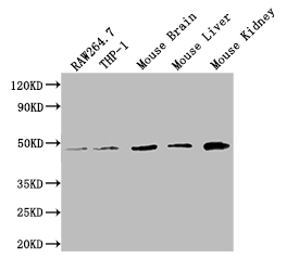
Western Blot Positive WB detected in: RAW264.7 whole cell lysate, THP-1 whole cell lysate, Mouse Brain tissue lysate, Mouse Liver tissue lysate, Mouse Kidney tissue lysate All lanes: WDFY1 antibody at 1:2000 Secondary Goat polyclonal to rabbit IgG at 1/50000 dilution Predicted band size: 47 kDa Observed band size: 47 kDa
Western Blot Positive WB detected in: RAW264.7 whole cell lysate, THP-1 whole cell lysate, Mouse Brain tissue lysate, Mouse Liver tissue lysate, Mouse Kidney tissue lysate All lanes: WDFY1 antibody at 1:2000 Secondary Goat polyclonal to rabbit IgG at 1/50000 dilution Predicted band size: 47 kDa Observed band size: 47 kDa
WDFY1 Antibody
CSB-PA816890LA01HU
ApplicationsWestern Blot, ELISA
Product group Antibodies
ReactivityHuman, Mouse
TargetWDFY1
Overview
- SupplierCusabio
- Product NameWDFY1 Antibody
- Delivery Days Customer20
- ApplicationsWestern Blot, ELISA
- CertificationResearch Use Only
- ClonalityPolyclonal
- ConjugateUnconjugated
- Gene ID57590
- Target nameWDFY1
- Target descriptionWD repeat and FYVE domain containing 1
- Target synonymsFENS-1, FENS1, WDF1, ZFYVE17, WD repeat and FYVE domain-containing protein 1, FYVE domain-containing protein localized to endosomes 1, WD40- and FYVE domain-containing protein 1, phosphoinositide-binding protein SR1, zinc finger FYVE domain-containing protein 17
- HostRabbit
- IsotypeIgG
- Protein IDQ8IWB7
- Protein NameWD repeat and FYVE domain-containing protein 1
- Scientific DescriptionPositively regulates TLR3- and TLR4-mediated signaling pathways by bridging the interaction between TLR3 or TLR4 and TICAM1. Promotes TLR3/4 ligand-induced activation of transcription factors IRF3 and NF-kappa-B, as well as the production of IFN-beta and inflammatory cytokines (PubMed:25736436).
- ReactivityHuman, Mouse
- Storage Instruction-20°C or -80°C
- UNSPSC41116161
Related products

Product group Antibodies
Anti-FENS1/WDFY1 Antibody Picoband(r)A10318-3-CARRIER-FREE
ApplicationsFlow Cytometry, Western Blot, ELISA
ReactivityHuman, Mouse, Rat
TargetWDFY1
- SizePrice

Product group Antibodies

Goat anti-FENS1 / WDFY1EB06341
ApplicationsELISA
ReactivityHuman, Mouse, Rat
TargetWDFY1
- SizePrice

Product group Antibodies
Anti-WDFY1 AntibodyHPA050603
ApplicationsWestern Blot, ImmunoHistoChemistry
ReactivityHuman
TargetWDFY1
- SizePrice
Product group Antibodies

WDFY1 AntibodyLS-C501305
ApplicationsWestern Blot, ELISA
ReactivityHuman, Mouse
TargetWDFY1
- SizePrice
Product group Antibodies
Wdfy1 Polyclonal AntibodyCAC09470
ApplicationsWestern Blot, ELISA
ReactivityMouse
TargetWDFY1
- SizePrice
Product group Antibodies
WDFY1 AntibodyPACO49234
ApplicationsWestern Blot, ELISA
ReactivityHuman, Mouse
TargetWDFY1
- SizePrice
![Non-transfected (–) and transfected (+) 293T whole cell extracts (30 μg) were separated by 10% SDS-PAGE, and the membrane was blotted with WDFY1+WDFY2 antibody [HL1989] (GTX637890) diluted at 1:5000. The HRP-conjugated anti-rabbit IgG antibody (GTX213110-01) was used to detect the primary antibody.](https://www.genetex.com/upload/website/prouct_img/normal/GTX637890/GTX637890_T-44872_20240112_WB_multiple_B_24011618_608.webp)
Product group Antibodies
WDFY1 + WDFY2 antibody [HL1989]GTX637890
ApplicationsWestern Blot
ReactivityHuman
TargetWDFY1
- SizePrice
Product group Antibodies
FENS1 Polyclonal AntibodyBS-13169R
ApplicationsFlow Cytometry, ImmunoFluorescence, ELISA, ImmunoCytoChemistry, ImmunoHistoChemistry, ImmunoHistoChemistry Frozen, ImmunoHistoChemistry Paraffin
ReactivityBovine, Canine, Equine, Human, Mouse, Porcine, Rabbit, Rat
TargetWDFY1
- SizePrice