


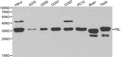
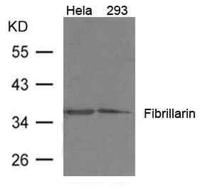
Figure 1. Western blot analysis of Fibrillarin/FBL using anti-Fibrillarin/FBL antibody (A03178-1). Electrophoresis was performed on a 5-20% SDS-PAGE gel at 70V (Stacking gel) / 90V (Resolving gel) for 2-3 hours. The sample well of each lane was loaded with 30 ug of sample under reducing conditions. Lane 1: monkey COS-7 whole cell lysates, Lane 2: human HEK293 whole cell lysates, Lane 3: human A375 whole cell lysates, Lane 4: human HepG2 whole cell lysates. After electrophoresis, proteins were transferred to a nitrocellulose membrane at 150 mA for 50-90 minutes. Blocked the membrane with 5% non-fat milk/TBS for 1.5 hour at RT. The membrane was incubated with rabbit anti-Fibrillarin/FBL antigen affinity purified polyclonal antibody (Catalog # A03178-1) at 0.25 microg/mL overnight at 4°C, then washed with TBS-0.1%Tween 3 times with 5 minutes each and probed with a goat anti-rabbit IgG-HRP secondary antibody at a dilution of 1:5000 for 1.5 hour at RT. The signal is developed using an Enhanced Chemiluminescent detection (ECL) kit (Catalog # EK1002) with Tanon 5200 system. A specific band was detected for Fibrillarin/FBL at approximately 37 kDa. The expected band size for Fibrillarin/FBL is at 37 kDa.
. Fibrillarin/FBL was detected in an immunocytochemical section of A549 cells. Enzyme antigen retrieval was performed using IHC enzyme antigen retrieval reagent (AR0022) for 15 mins. The cells were blocked with 10% goat serum. And then incubated with 5 microg/mL rabbit anti-Fibrillarin/FBL Antibody (A03178-1) overnight at 4°C. DyLight®594 Conjugated Goat Anti-Rabbit IgG (BA1142) was used as secondary antibody at 1:100 dilution and incubated for 30 minutes at 37°C. The tissue section was developed using Phalloidin-iFluor 488 Conjugated. Visualize using a fluorescence microscope and filter sets appropriate for the label used.")
. Overlay histogram showing U20S cells stained with A03178-1 (Blue line). To facilitate intracellular staining, cells were fixed with 4% paraformaldehyde and permeabilized with permeabilization buffer. The cells were blocked with 10% normal goat serum. And then incubated with rabbit anti-Fibrillarin/FBL Antibody (A03178-1, 1 microg/1x106 cells) for 30 min at 20°C. DyLight®488 conjugated goat anti-rabbit IgG (BA1127, 5-10 microg/1x106 cells) was used as secondary antibody for 30 minutes at 20°C. Isotype control antibody (Green line) was rabbit IgG (1 microg/1x106) used under the same conditions. Unlabelled sample without incubation with primary antibody and secondary antibody (Red line) was used as a blank control.")
Figure 1. Western blot analysis of Fibrillarin/FBL using anti-Fibrillarin/FBL antibody (A03178-1). Electrophoresis was performed on a 5-20% SDS-PAGE gel at 70V (Stacking gel) / 90V (Resolving gel) for 2-3 hours. The sample well of each lane was loaded with 30 ug of sample under reducing conditions. Lane 1: monkey COS-7 whole cell lysates, Lane 2: human HEK293 whole cell lysates, Lane 3: human A375 whole cell lysates, Lane 4: human HepG2 whole cell lysates. After electrophoresis, proteins were transferred to a nitrocellulose membrane at 150 mA for 50-90 minutes. Blocked the membrane with 5% non-fat milk/TBS for 1.5 hour at RT. The membrane was incubated with rabbit anti-Fibrillarin/FBL antigen affinity purified polyclonal antibody (Catalog # A03178-1) at 0.25 microg/mL overnight at 4°C, then washed with TBS-0.1%Tween 3 times with 5 minutes each and probed with a goat anti-rabbit IgG-HRP secondary antibody at a dilution of 1:5000 for 1.5 hour at RT. The signal is developed using an Enhanced Chemiluminescent detection (ECL) kit (Catalog # EK1002) with Tanon 5200 system. A specific band was detected for Fibrillarin/FBL at approximately 37 kDa. The expected band size for Fibrillarin/FBL is at 37 kDa.
Anti-Fibrillarin/FBL Antibody Picoband(r)
A03178-1-CARRIER-FREE
ApplicationsFlow Cytometry, ImmunoFluorescence, Western Blot, ELISA, ImmunoCytoChemistry
Product group Antibodies
ReactivityHuman, Monkey
TargetFBL
Overview
- SupplierBoster Bio
- Product NameAnti-Fibrillarin/FBL Antibody Picoband(r)
- Delivery Days Customer9
- ApplicationsFlow Cytometry, ImmunoFluorescence, Western Blot, ELISA, ImmunoCytoChemistry
- CertificationResearch Use Only
- ClonalityPolyclonal
- Concentration500 ug/ml
- Gene ID2091
- Target nameFBL
- Target descriptionfibrillarin
- Target synonymsFIB, FLRN, Nop1, RNU3IP1, rRNA 2'-O-methyltransferase fibrillarin, 34 kDa nucleolar scleroderma antigen, 34-kD nucleolar scleroderma antigen, RNA, U3 small nucleolar interacting protein 1, U6 snRNA 2'-O-methyltransferase fibrillarin, histone-glutamine methyltransferase
- HostRabbit
- IsotypeIgG
- Protein IDP22087
- Protein NamerRNA 2'-O-methyltransferase fibrillarin
- Scientific DescriptionBoster Bio Anti-Fibrillarin/FBL Antibody Picoband® catalog # A03178-1. Tested in ELISA, Flow Cytometry, IF, ICC, WB applications. This antibody reacts with Human, Monkey. The brand Picoband indicates this is a premium antibody that guarantees superior quality, high affinity, and strong signals with minimal background in Western blot applications. Only our best-performing antibodies are designated as Picoband, ensuring unmatched performance.
- ReactivityHuman, Monkey
- Storage Instruction-20°C,2°C to 8°C
- UNSPSC12352203
Related products
Product group Antibodies
Anti-FBL AntibodyA29417
ApplicationsImmunoFluorescence, ImmunoPrecipitation, Western Blot, ImmunoHistoChemistry, Other Application
ReactivityHuman, Mouse, Rat
- SizePrice
Product group Antibodies
Anti-FBL Antibody144-60358
ApplicationsImmunoFluorescence, ImmunoPrecipitation, Western Blot, ImmunoHistoChemistry
ReactivityHuman, Monkey, Mouse, Rat
TargetFBL
- SizePrice
Product group Antibodies

FBL / FIB / Fibrillarin AntibodyLS-C748539
ApplicationsImmunoFluorescence, ImmunoPrecipitation, Western Blot, ImmunoHistoChemistry
ReactivityHuman, Monkey, Mouse, Rat
TargetFBL
- SizePrice
Product group Antibodies
Fibrillarin Polyclonal AntibodyBS-3998R
ApplicationsWestern Blot, ELISA
ReactivityBovine, Canine, Human, Mouse, Porcine, Rat
TargetFBL
- SizePrice
Product group Antibodies

Goat anti-Fibrillarin / FBLEB11640
ApplicationsWestern Blot, ELISA
ReactivityBovine, Canine, Human, Mouse, Porcine, Rat
TargetFBL
- SizePrice
Product group Antibodies
ApplicationsImmunoPrecipitation, Western Blot, ImmunoCytoChemistry, ImmunoHistoChemistry
ReactivityMouse, Rat
TargetFBL
- SizePrice
Product group Antibodies
FBL AntibodyCSB-PA919400
ApplicationsWestern Blot, ELISA
ReactivityHuman, Mouse, Rat
TargetFBL
- SizePrice

Product group Antibodies
Fibrillarin antibodyGTX101807
ApplicationsImmunoFluorescence, ImmunoPrecipitation, Western Blot, ImmunoCytoChemistry
ReactivityHuman, Mouse, Rat
TargetFBL
- SizePrice

Product group Antibodies
Anti-FBL AntibodyHPA049546
ApplicationsImmunoCytoChemistry
ReactivityHuman
TargetFBL
- SizePrice