


Immunofluorescent staining of human cell line A-431 shows localization to nucleoplasm.

Immunofluorescent staining of human cell line A-431 shows localization to nucleoplasm.
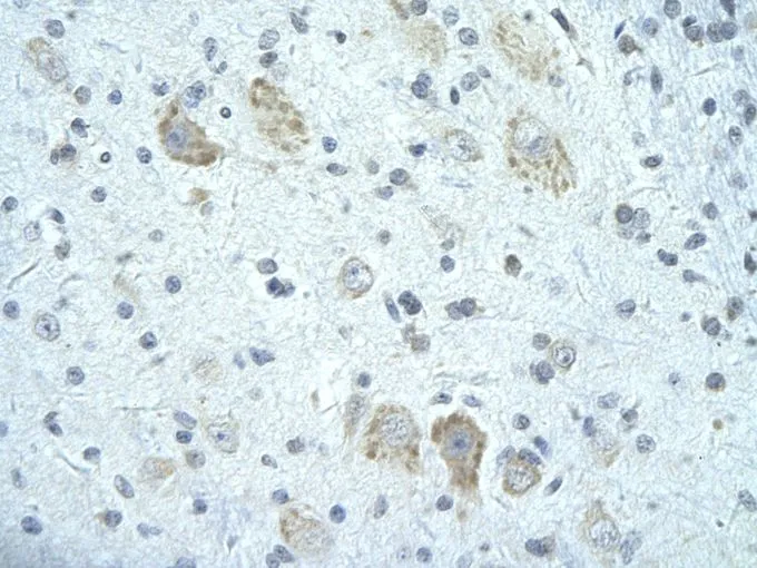
Anti-FIP1L1 Antibody
HPA058202
ApplicationsWestern Blot, ImmunoCytoChemistry
Product group Antibodies
ReactivityHuman
TargetFIP1L1
Overview
- SupplierAtlas Antibodies
- Product NameAnti-FIP1L1 Antibody
- Delivery Days Customer4
- ApplicationsWestern Blot, ImmunoCytoChemistry
- CertificationResearch Use Only
- ClonalityPolyclonal
- ConjugateUnconjugated
- Gene ID81608
- Target nameFIP1L1
- Target descriptionfactor interacting with PAPOLA and CPSF1
- Target synonymsFIP1, Rhe, hFip1, pre-mRNA 3'-end-processing factor FIP1, FIP1 like 1, FIP1-like 1 protein, FIP1L1 cleavage and polyadenylation specific factor subunit, factor interacting with PAP, rearranged in hypereosinophilia
- HostRabbit
- IsotypeIgG
- Protein IDQ6UN15
- Protein NamePre-mRNA 3'-end-processing factor FIP1
- Scientific DescriptionRecombinant Protein Epitope Signature Tag (PrEST) antigen sequence
- ReactivityHuman
- Storage Instruction-20°C,2°C to 8°C
- UNSPSC41116161
Datasheet
MSDS
Related products
Product group Antibodies
Anti-FIP1L1 AntibodyA31903
ApplicationsImmunoFluorescence, Western Blot, ImmunoHistoChemistry
ReactivityHuman, Mouse, Rat
- SizePrice

Product group Antibodies
Anti-FIP1L1 Antibody Picoband(r)A02452-1-CARRIER-FREE
ApplicationsFlow Cytometry, ImmunoFluorescence, Western Blot, ELISA, ImmunoCytoChemistry, ImmunoHistoChemistry
ReactivityHuman, Mouse, Rat
TargetFIP1L1
- SizePrice
Product group Antibodies
Anti-FIP1L1 Antibody144-07138
ApplicationsImmunoFluorescence, Western Blot, ImmunoHistoChemistry
ReactivityHuman, Mouse, Rat
TargetFIP1L1
- SizePrice
Product group Antibodies
FIP1L1 Polyclonal AntibodyBS-13173R
ApplicationsImmunoFluorescence, Western Blot, ELISA, ImmunoCytoChemistry, ImmunoHistoChemistry, ImmunoHistoChemistry Frozen, ImmunoHistoChemistry Paraffin
ReactivityBovine, Canine, Chicken, Equine, Human, Mouse, Porcine, Rabbit, Rat, Sheep
TargetFIP1L1
- SizePrice
Product group Antibodies

HFip1 / FIP1L1 AntibodyLS-C346244
ApplicationsImmunoFluorescence, Western Blot, ImmunoHistoChemistry
ReactivityHuman, Mouse, Rat
TargetFIP1L1
- SizePrice
Product group Antibodies
FIP1L1 antibody, C-termGTX47245
ApplicationsWestern Blot, ImmunoHistoChemistry, ImmunoHistoChemistry Paraffin
ReactivityHuman
TargetFIP1L1
- SizePrice

Product group Antibodies
Anti-FIP1L1 AntibodyHPA037475
ApplicationsWestern Blot, ImmunoCytoChemistry, ImmunoHistoChemistry
ReactivityHuman
TargetFIP1L1
- SizePrice
Product group Antibodies
Anti-FIP1L1 AntibodyHPA037475
ApplicationsWestern Blot, ImmunoCytoChemistry, ImmunoHistoChemistry
ReactivityHuman
TargetFIP1L1
- SizePrice