

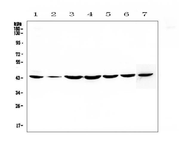
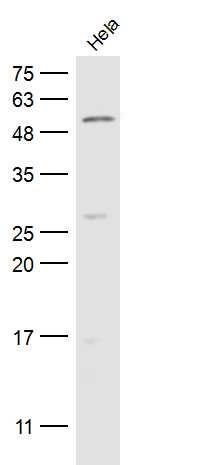
Figure 1. Western blot analysis of FLIP using anti-FLIP antibody (A01295-1). Electrophoresis was performed on a 5-20% SDS-PAGE gel at 70V (Stacking gel) / 90V (Resolving gel) for 2-3 hours. The sample well of each lane was loaded with 50ug of sample under reducing conditions. Lane 1: human Hela whole cell lysates, Lane 2: human placenta tissue lysates, Lane 3: human COLO-320 whole cell lysates, Lane 4: human PANC-1 whole cell lysates, Lane 5: human HepG2 whole cell lysates, Lane 6: human MDA-MB-231 whole cell lysates, Lane 7: mouse NIH3T3 whole cell lysates. After Electrophoresis, proteins were transferred to a Nitrocellulose membrane at 150mA for 50-90 minutes. Blocked the membrane with 5% Non-fat Milk/ TBS for 1.5 hour at RT. The membrane was incubated with rabbit anti-FLIP antigen affinity purified polyclonal antibody (Catalog # A01295-1) at 0.5 microg/mL overnight at 4°C, then washed with TBS-0.1%Tween 3 times with 5 minutes each and probed with a goat anti-rabbit IgG-HRP secondary antibody at a dilution of 1:10000 for 1.5 hour at RT. The signal is developed using an Enhanced Chemiluminescent detection (ECL) kit (Catalog # EK1002) with Tanon 5200 system. A specific band was detected for FLIP at approximately 43KD. The expected band size for FLIP is at 55KD.
. FLIP was detected in paraffin-embedded section of mouse small intestine tissue. Heat mediated antigen retrieval was performed in citrate buffer (pH6, epitope retrieval solution) for 20 mins. The tissue section was blocked with 10% goat serum. The tissue section was then incubated with 2microg/ml rabbit anti-FLIP Antibody (A01295-1) overnight at 4°C. Biotinylated goat anti-rabbit IgG was used as secondary antibody and incubated for 30 minutes at 37°C. The tissue section was developed using Strepavidin-Biotin-Complex (SABC)(Catalog # SA1022) with DAB as the chromogen.")
. FLIP was detected in paraffin-embedded section of rat small intestine tissue. Heat mediated antigen retrieval was performed in citrate buffer (pH6, epitope retrieval solution) for 20 mins. The tissue section was blocked with 10% goat serum. The tissue section was then incubated with 2microg/ml rabbit anti-FLIP Antibody (A01295-1) overnight at 4°C. Biotinylated goat anti-rabbit IgG was used as secondary antibody and incubated for 30 minutes at 37°C. The tissue section was developed using Strepavidin-Biotin-Complex (SABC)(Catalog # SA1022) with DAB as the chromogen.")
Figure 1. Western blot analysis of FLIP using anti-FLIP antibody (A01295-1). Electrophoresis was performed on a 5-20% SDS-PAGE gel at 70V (Stacking gel) / 90V (Resolving gel) for 2-3 hours. The sample well of each lane was loaded with 50ug of sample under reducing conditions. Lane 1: human Hela whole cell lysates, Lane 2: human placenta tissue lysates, Lane 3: human COLO-320 whole cell lysates, Lane 4: human PANC-1 whole cell lysates, Lane 5: human HepG2 whole cell lysates, Lane 6: human MDA-MB-231 whole cell lysates, Lane 7: mouse NIH3T3 whole cell lysates. After Electrophoresis, proteins were transferred to a Nitrocellulose membrane at 150mA for 50-90 minutes. Blocked the membrane with 5% Non-fat Milk/ TBS for 1.5 hour at RT. The membrane was incubated with rabbit anti-FLIP antigen affinity purified polyclonal antibody (Catalog # A01295-1) at 0.5 microg/mL overnight at 4°C, then washed with TBS-0.1%Tween 3 times with 5 minutes each and probed with a goat anti-rabbit IgG-HRP secondary antibody at a dilution of 1:10000 for 1.5 hour at RT. The signal is developed using an Enhanced Chemiluminescent detection (ECL) kit (Catalog # EK1002) with Tanon 5200 system. A specific band was detected for FLIP at approximately 43KD. The expected band size for FLIP is at 55KD.
Anti-FLIP/CFLAR Antibody Picoband(r)
A01295-1-CARRIER-FREE
ApplicationsWestern Blot, ELISA, ImmunoHistoChemistry
Product group Antibodies
ReactivityHuman, Mouse, Rat
TargetCFLAR
Overview
- SupplierBoster Bio
- Product NameAnti-FLIP/CFLAR Antibody Picoband(r)
- Delivery Days Customer9
- ApplicationsWestern Blot, ELISA, ImmunoHistoChemistry
- CertificationResearch Use Only
- ClonalityPolyclonal
- Concentration500 ug/ml
- Gene ID8837
- Target nameCFLAR
- Target descriptionCASP8 and FADD like apoptosis regulator
- Target synonymsCASH, CASP8AP1, CLARP, Casper, FLAME, FLAME-1, FLAME1, FLIP, I-FLICE, MRIT, c-FLIP, c-FLIPL, c-FLIPR, c-FLIPS, cFLIP, CASP8 and FADD-like apoptosis regulator, FADD-like antiapoptotic molecule 1, MACH-related inducer of toxicity, caspase homolog, caspase-eight-related protein, caspase-like apoptosis regulatory protein, caspase-related inducer of apoptosis, cellular FLICE-like inhibitory protein, inhibitor of FLICE, testis secretory sperm-binding protein Li 233m, usurpin beta
- HostRabbit
- IsotypeIgG
- Protein IDO15519
- Protein NameCASP8 and FADD-like apoptosis regulator
- Scientific DescriptionBoster Bio Anti-FLIP/CFLAR Antibody Picoband® catalog # A01295-1. Tested in ELISA, IHC, WB applications. This antibody reacts with Human, Mouse, Rat. The brand Picoband indicates this is a premium antibody that guarantees superior quality, high affinity, and strong signals with minimal background in Western blot applications. Only our best-performing antibodies are designated as Picoband, ensuring unmatched performance.
- ReactivityHuman, Mouse, Rat
- Storage Instruction-20°C,2°C to 8°C
- UNSPSC12352203
Related products
Product group Antibodies
CFLAR AntibodyCSB-PA004981
ApplicationsWestern Blot, ELISA
ReactivityHuman
TargetCFLAR
- SizePrice
Product group Antibodies
Anti-CFLAR AntibodyA101156
ApplicationsWestern Blot, ELISA
ReactivityHuman
- SizePrice

Product group Antibodies
Anti-CFLAR AntibodyHPA019044
ApplicationsImmunoCytoChemistry, ImmunoHistoChemistry
ReactivityHuman
TargetCFLAR
- SizePrice
Product group Antibodies

CFLAR / FLIP AntibodyLS-C332165
ApplicationsWestern Blot, ImmunoHistoChemistry
ReactivityHuman, Mouse, Rat
TargetCFLAR
- SizePrice
Product group Antibodies
anti-FLIP, mAb (Dave-2)AG-20B-0005
ApplicationsImmunoPrecipitation, Western Blot
ReactivityHuman, Mouse
TargetCFLAR
- SizePrice
Product group Antibodies
Cflar Polyclonal AntibodyCAC08309
ApplicationsImmunoFluorescence, Western Blot, ELISA, ImmunoHistoChemistry
TargetCFLAR
- SizePrice
![Whole cell extract (30 μg) was separated by 10% SDS-PAGE, and the membranes were blotted with FLIP antibody [N1C1] (GTX113047) diluted at 1:500 and competitor's antibody (sc-8347) diluted at 1:200. The HRP-conjugated anti-rabbit IgG antibody (GTX213110-01) was used to detect the primary antibody.](https://www.genetex.com/upload/website/prouct_img/normal/GTX113047/GTX113047_40394_20170825_WB_competitor_watermark_w_23060500_896.webp)
Product group Antibodies
FLIP antibody [N1C1]GTX113047
ApplicationsWestern Blot, ImmunoHistoChemistry, ImmunoHistoChemistry Paraffin
ReactivityHuman
TargetCFLAR
- SizePrice
Product group Antibodies
FLIP Polyclonal AntibodyBS-0119R
ApplicationsFlow Cytometry, ImmunoFluorescence, Western Blot, ELISA, ImmunoCytoChemistry, ImmunoHistoChemistry, ImmunoHistoChemistry Frozen, ImmunoHistoChemistry Paraffin
ReactivityBovine, Canine, Human, Mouse, Porcine, Rabbit, Rat
TargetCFLAR
- SizePrice
Product group Antibodies
Anti-CFLAR Antibody144-65545
ApplicationsWestern Blot, ImmunoHistoChemistry
ReactivityHuman, Mouse, Rat
TargetCFLAR
- SizePrice